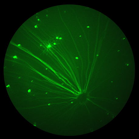
In their paper, “Suppression of Choroidal Neovascularization by AAV-Based Dual-Acting Antiangiogenic Gene Therapy,” Askou et al develop an adeno-associated virus (AAV) treatment for age-related macular degeneration. Beautiful fluorescent fundoscopy performed with the Phoenix MICRON® validated the success of the subretinal AAV injection, while precise choroidal neovascularization (CNV) induced by Phoenix laser burns confirmed that the […]
15.02
2019
Corneal Thickness Analysis using OCT
Corneal images taken with the Phoenix Micron IV OCT used for thickness analysis King et al, a consortium of researchers at a range of institutions, recently used the Phoenix Micron IV OCT to examine corneal thickness in their article, “Genomic locus modulating corneal thickness in the mouse identifies POU6F2 as a potential risk of developing […]
17.01
2019
Characterizing a mutant rat strain with the Phoenix Micron OCT and Ganzfeld ERG
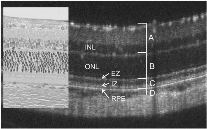
Monai et al characterized the longitudinal retinal degeneration of a rat model of retinitis pigmentosa using the Phoenix Micron OCT to examine retinal layers in live rats and the full field Ganzfeld ERG to test function. The rats have one of the mutations, P23H, that cause retinitis pigmentosa in humans, and are specifically a very […]
16.11
2018
Fundus and OCT imaging shows that smart phone-like blue light exposure leads to retinal disruption in rats
Researchers Lin et al at Taipei Medical University shared an alarming finding that blue light, similar to that emitted by smart phones, can lead to retinal disruption in rats. They used the Phoenix-Micron’s Phoenix MICRON® fundus camera and the image-guided OCT to demonstrate blood vessel leakage and retinal thinning after intermittent blue light exposure. Lin […]
11.07
2018
Phoenix’s anterior chamber OCT evaluates corneal wound healing in guinea pigs
McDaniels et al at the US Army Institute of Surgical Research used the Phoenix anterior chamber OCT to evaluate the safety of an ocular wound chamber after a corneal injury in guinea pigs. The research group developed an ocular wound chamber that encases the eye and allows for controlled delivery of therapeutics and eye lubricant. […]
12.06
2018
Phoenix OCT reveals detailed images of retinal and anterior chamber infiltrates in a uveitis mouse model
In their article “Multimodal analysis of ocular inflammation using the endotoxin-induced uveitis mouse model,” Chu et al used images captured with the Phoenix OCT of the retina and anterior segment to correlate infiltrate level with neutrophil count during uveitis. To develop objective multimodal methods of analyzing infected eyes, they also examined flow cytometry of infiltrates, […]


