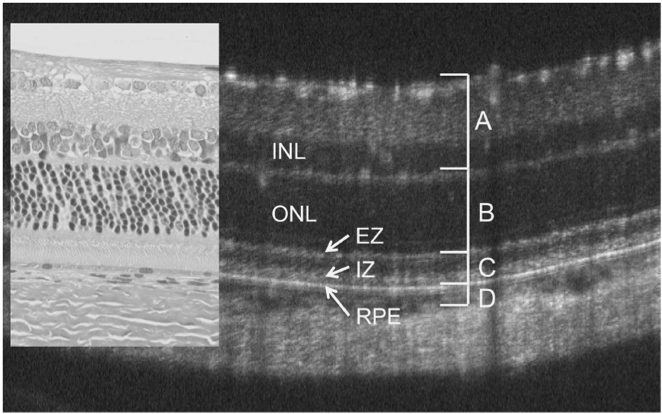
In the paper “The impact of obesity‑related raised intracranial pressure in rodents” published in Scientific Reports journal, Westgate and collaborators used the Phoenix MICRON® image-guided OCT system, along with the Phoenix MICRON® Insight software, in the measurement of the retinal anatomical changes caused by intracranial hypertension in obese rats. The Problem: Intracranial pressure (ICP) measures the […]
29.11
2021
Phoenix MICRON Spins out of Phoenix Technology Group to Better Serve Eye and Eye-Brain Researchers Globally
Bend, OR, USA, November 29, 2021 — The newly formed company, Phoenix-Micron, Inc., announced today it has completed the spin-out of the Phoenix MICRON® imaging platform from Phoenix Technology Group. This move is designed to increase focus and innovation in products designed to serve the eye and eye-brain research community. The new company, Phoenix-Micron, Inc. […]
21.07
2020
Caspase-9 inhibiting eyedrops rescue physiological and functional retinal vein occlusion damage shown with Phoenix MICRON®, OCT, and focal ERG
In a recent well written, compelling article published in Nature Communications, “Endothelial activation of caspase-9 promotes neurovascular injury in retinal vein occlusion,” Avrutsky et al show that caspase-9 inhibition is a promising treatment for retinal vein occlusion. Retinal vein occlusion models hypoxic-ischemic neurovascular damage and is the second leading cause of blindness in working-age adults. […]
23.06
2020
Targeting VEGF164 in Müller cells may be useful to treat retinopathy of prematurity
In Nature’s Scientific Reports, Becker et al use the Phoenix MICRON® IV, OCT, and focal ERG to assess the therapeutic value of knocking down a splice variant of VEGF in Müller cells in a model of Retinopathy of Prematurity (ROP). ROP is characterized by delayed vascularization of the retina after disrupted oxygen levels, followed by […]
15.02
2019
Corneal Thickness Analysis using OCT
Corneal images taken with the Phoenix Micron IV OCT used for thickness analysis King et al, a consortium of researchers at a range of institutions, recently used the Phoenix Micron IV OCT to examine corneal thickness in their article, “Genomic locus modulating corneal thickness in the mouse identifies POU6F2 as a potential risk of developing […]
17.01
2019
Characterizing a mutant rat strain with the Phoenix Micron OCT and Ganzfeld ERG
Monai et al characterized the longitudinal retinal degeneration of a rat model of retinitis pigmentosa using the Phoenix Micron OCT to examine retinal layers in live rats and the full field Ganzfeld ERG to test function. The rats have one of the mutations, P23H, that cause retinitis pigmentosa in humans, and are specifically a very […]