Monai et al characterized the longitudinal retinal degeneration of a rat model of retinitis pigmentosa using the Phoenix Micron OCT to examine retinal layers in live rats and the full field Ganzfeld ERG to test function. The rats have one of the mutations, P23H, that cause retinitis pigmentosa in humans, and are specifically a very slow degeneration model (line 2) that has not been thoroughly characterized before.
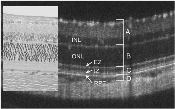
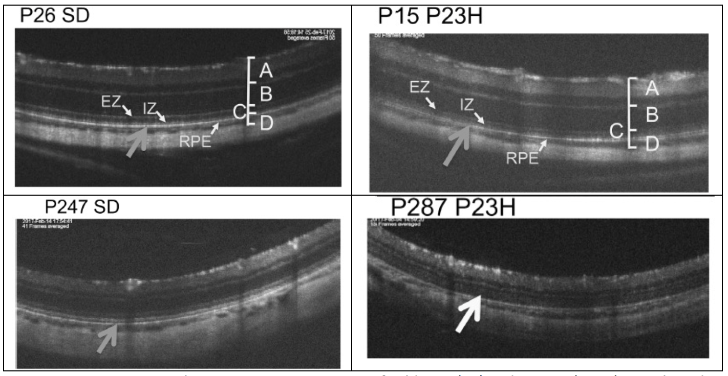
Figure 1 shows the strong correlation of the Phoenix OCT images with a histological section of a wild type rat retina, proving the OCT is a powerful tool for live, longitudinal retinal health analysis. The P23H rats had normal retinae up to about P43, but the photoreceptor inner and outer segment layer became diffusely hyper-reflective after P43 (Fig 2). The researchers continued OCT examination up to day P287, which demonstrates the power of longitudinal studies. Electron microscopy confirmed that the OCT changes were due to disruption in the arrangement of the outer segment discs.
Using the Phoenix Insight semi-automated segmentation software, Monai et al found that the outer plexiform and outer nuclear layers rapidly thinned after P71. The inner and outer segment layer thinning was more gradual and was only significantly thinner than the wild type rats after P237.
The researchers used the Phoenix full field Ganzfeld ERG system to test the function of the P23H mutant rats. Both the a waves (a measure of photoreceptor function) and b waves (bipolar cell function) decreased with age for the wild type and mutant rats. However, at P99, the a and b waves of the P23H rats were significantly reduced compared to wild type rats of the same age (Fig 3). Statistical analysis revealed that the P23H a and b wave amplitude was correlated with the outer nuclear and outer plexiform layers.
Reference: Monai N, Yamauchi K, Tanabu R, Gonome T, Ishiguro S-i, Nakazawa M. (2018). Characterization of photoreceptor degeneration in the rhodopsin P23H transgenic rat line 2 using optical coherence tomography. PLoS ONE 13(3):e0193778. https://doi.org/10.1371/journal.pone.0193778


