For astronauts gazing out at the vastness of space, their eyes are not just windows to the cosmos but sensitive and vital instruments that must endure the challenging environment of life beyond Earth’s atmosphere. Although pressure suits, capsules, and space stations provide critical protection, astronauts are constantly exposed to extraterrestrial forces that impact various organ […]
10.07
2024
Tau Protein Modulation Impacts Retinal Neuron Survival in Glaucoma
Glaucoma, a leading cause of irreversible vision loss, is characterized by progressive damage to retinal ganglion cells (RGCs) and the optic nerve, often associated with increased intraocular pressure (IOP). While previous studies have implicated Tau protein expression and phosphorylation changes in other neurodegenerative diseases such as Alzheimer’s disease, Parkinson’s disease and glaucoma, the causative role […]
08.04
2024
MICRON Imaging Systems: Powering 900 Scientific Publications
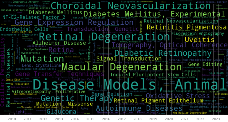
The ability to see the world around us is a gift that’s easy to take for granted, until it’s threatened by the onset of a devastating eye disease. For those facing the reality of vision loss, ophthalmic research represents a beacon of hope, offering the promise of new treatments and therapies to preserve or restore […]
01.02
2024
Phoenix-Micron, Inc. Announces Exclusive North American Distribution of Striatech Products
Phoenix extends its product offerings in North America to include behavioral assays to evaluate the visual abilities of rodents Bend, OR, USA, January 25, 2024 – Phoenix-Micron, Inc. (“Phoenix”), recognized globally for its leadership in in vivo ophthalmic imaging of small animals, is proud to unveil its latest collaboration as the exclusive North American […]
28.02
2022
Analysis of imaging, structure and function using Phoenix MICRON™ modalities expand the understanding of ocular features of Down Syndrome in mouse models
In their paper “Quantitative Analysis of Retinal Structure and Function in Two Chromosomally Altered Mouse Models of Down Syndrome”, researchers Victorino, Scott-McKean, et al leveraged the multi-modality capabilities of the Phoenix MICRON™ retinal imaging platform, to produce an image-rich research paper looking at the ocular features of Down Syndrome in two mouse models; Ts65Dn and […]
29.11
2021
Phoenix MICRON Spins out of Phoenix Technology Group to Better Serve Eye and Eye-Brain Researchers Globally
Bend, OR, USA, November 29, 2021 — The newly formed company, Phoenix-Micron, Inc., announced today it has completed the spin-out of the Phoenix MICRON® imaging platform from Phoenix Technology Group. This move is designed to increase focus and innovation in products designed to serve the eye and eye-brain research community. The new company, Phoenix-Micron, Inc. […]


