In their 2017 article, “Effect of subretinal injection on retinal structure and function in a rat oxygen-induced retinopathy model,” Becker et al used the Phoenix MICRON® IV fundus camera, Phoenix MICRON® OCT2 and corresponding layer analysis software Insight 2D, and the Phoenix MICRON® focal ERG to find that subretinal injection of saline or even introduction […]
24.06
2021
Promising treatment for retinal inflammation studied with the Phoenix MICRON® OCT and exclusive analysis software Insight 2D
In their paper, “Connexin43 Mimetic Peptide Improves Retinal Function and Reduces Inflammation in a Light-Damaged Albino Rat Model,” Guo et al promoted neuroretinal survival in a light damage paradigm by blocking the Connexin43 hemichannels. Using the Phoenix MICRON® OCT to examine the structure of the retina at precise retinal locations as shown by the bright […]
27.01
2021
Stunning fundus images of GFP-positive cells demonstrate new intravitreal injection technique
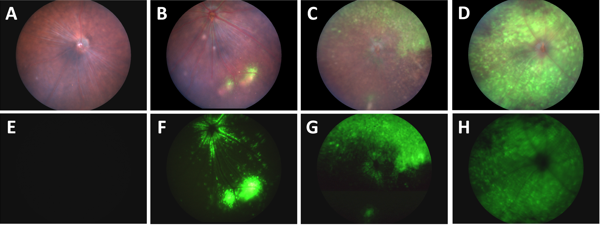
In their article, “A Novel Method Combining Vitreous Aspiration and Intravitreal AAV2/8 Injection Results in Retina-Wide Transduction in Adult Mice,” Da Costa et al use the Phoenix MICRON® III imaging platform to take stunning images demonstrating the success of their novel intravitreal injection technique. Gene therapy is a promising treatment option of genetic retinopathies—adeno-associated viruses […]
28.10
2020
A year-long longitudinal pattern dystrophy fundus study with the Phoenix MICRON® IV imaging platform
In their 2019 paper, “Novel molecular mechanisms for Prph2‐associated pattern dystrophy,” Chakraborty et al use the Phoenix MICRON® IV retinal imaging platform to longitudinally study the effect of a very specific mutation affecting the Peripherin 2 protein. Peripherin 2 is a protein in rods and cones which, if mutated, can lead to retinitis pigmentosa, cone-rod […]
29.09
2020
RPE mutations lead to retinal hypopigmentation, vasculature changes, and decreased function
In their paper, “The microphthalmia-associated transcription factor (Mitf) gene and its role in regulating eye function,” García-Llorca et al use the Phoenix MICRON® IV to examine the outer eye appearance, retinal pigmentation, and retinal vasculature through fluorescein angiography to study several different mouse mutants. Combined with electroretinography and histology, the fundus images tell a story […]