Perform segmentation and data analysis from your desk, not the imaging station. Lab space isn’t always the best place to work with your study images and data. Between PPE requirements, access restrictions, and competition for imaging time, the lab can slow down your analysis workflow. MICRON Review Station changes that. What It Does MICRON Review […]
13.11
2025
Advancing Geographic Atrophy Research with Image-Guided Laser Technology
In April 2025, we announced the MICRON Image-Guided 810 nm Laser system with the ability to induce geographic atrophy in rodents. Today I’m happy to announce that the system has started shipping to researchers, and the final product includes three laser modes: continuous wave, long pulse, and short pulse. In this blog post we provide […]
24.06
2025
Phoenix-Micron, Inc. is Releasing Glove Liners to Support Researchers With Rodent Bite Injury Solutions.
We are excited to release new glove liners to the MICRON eStore. We know that working with rodents in a lab environment comes with risks—bites and scratches or inadvertent needle pokes can lead to injury and contamination. Because of this, MICRON glove liners are engineered to provide an extra layer of protection to avoid rodent […]
12.09
2024
MICRON® Imaging Systems: Out of This World – Advancing Space Exploration
For astronauts gazing out at the vastness of space, their eyes are not just windows to the cosmos but sensitive and vital instruments that must endure the challenging environment of life beyond Earth’s atmosphere. Although pressure suits, capsules, and space stations provide critical protection, astronauts are constantly exposed to extraterrestrial forces that impact various organ […]
24.07
2024
Newly Discovered Eye-Brain Immune Connection Opens Doors for Novel Therapies
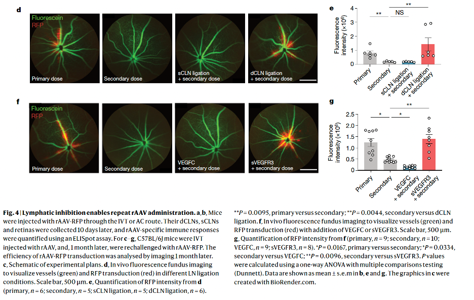
Researchers at Yale University have uncovered a previously unknown lymphatic drainage system connecting the back of the eye to the brain, potentially revolutionizing our understanding of eye-brain immunity and opening new avenues for treating central nervous system diseases. By studying the immune response to herpes simplex virus in the brain, the team led by Dr. […]
12.12
2023
Exploring the Intersection of Art and Retinal Research: A Journey of Discovery

Our latest blog post veers from the traditional focus on a singular research paper to bring you an enthralling narrative at the crossroads of progressive inherited retinal dystrophy (IRD), the frigid expanses of Antarctica, marathon endurance, the expressive world of art, and the transformative realm of retinal research. This story encompasses gene editing, animal models, […]


