Researchers at Henan Province Eye Hospital in China have made great strides in studying endotoxin-induced uveitis (EIU) by creating a novel EIU model in zebrafish. Uveitis is an ocular inflammation and one of the main causes of visual impairment, accounting for 10-15% of global blindness. Mice have typically been the research animal of choice however the time course can be long and the process is expensive therefore an ideal model for the disease remains to be seen. Zebrafish are smaller to house, have a faster life cycle, are easy to breed and, like mice, are compatible with gene-editing.
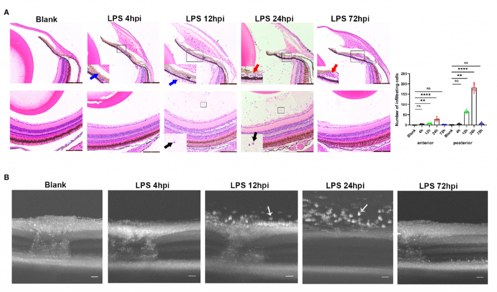
In their paper “A novel uveitis model induced by lipopolysaccharide in zebrafish” published in Frontiers of Immunologyin 2022, investigators Xiao Xiao et al successfully induced EIU in zebrafish eyes following an intravitreal injection oflipopolysaccharide (LPS). Longitudinal OCT imaging using the Phoenix MICRON® retinal imaging platform, beautifully documented the infiltration of cells as hyperreflective dots, with the onset of inflammation at 4 hours post injection (hpi), reaching its peak at 24 hpi, and then resolving at 72 hpi. Immunofluorescence and histology at the same time points matched the imaging series. It is notable that the effective LPS dose required to induce intraocular inflammation in zebrafish was 32 times higher than that of the mouse, while the rapid spontaneous resolution was consistent with the murine model. Inflammation was inhibited by immersing the zebrafish in prednisone.
This study demonstrated the feasibility of inducing, imaging and controlling EIU in zebrafish, creating promise of new pathways for research into gene-editing, screening and the development of novel drugs for this debilitating eye disease. For more details on this remarkable work, take a look at the original paper Xiao X, Liu Z, Su G, Liu H, Yin W, Guan Y, Jing S, Du L, Li F, Li N and Yang P (2022) A novel uveitis model induced by lipopolysaccharide in zebrafish.Front. Immunol. 13:1042849. doi: 10.3389/fimmu.2022.1042849