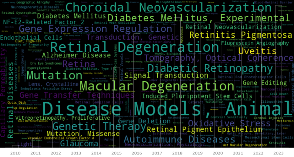
The ability to see the world around us is a gift that’s easy to take for granted, until it’s threatened by the onset of a devastating eye disease. For those facing the reality of vision loss, ophthalmic research represents a beacon of hope, offering the promise of new treatments and therapies to preserve or restore […]
25.03
2021
Therapeutic target for retinal degeneration studied with the Phoenix MICRON® retinal structure and function tools
In their article, “Effect of MMP-9 gene knockout on retinal vascular form and function,” George et al study the effect of knocking out a matrix protein in a mouse model of retinitis pigmentosa using the Phoenix MICRON® platform including OCT, and Ganzfeld ERG. The combination of the Phoenix MICRON® fundus images, OCT revealing the layers, […]
29.09
2020
RPE mutations lead to retinal hypopigmentation, vasculature changes, and decreased function
In their paper, “The microphthalmia-associated transcription factor (Mitf) gene and its role in regulating eye function,” García-Llorca et al use the Phoenix MICRON® IV to examine the outer eye appearance, retinal pigmentation, and retinal vasculature through fluorescein angiography to study several different mouse mutants. Combined with electroretinography and histology, the fundus images tell a story […]
17.01
2019
Characterizing a mutant rat strain with the Phoenix Micron OCT and Ganzfeld ERG
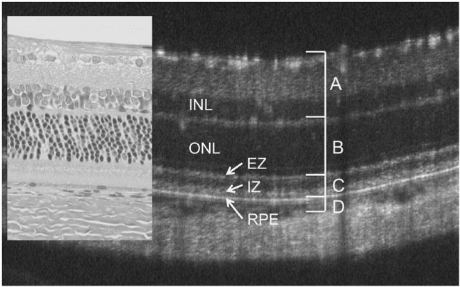
Monai et al characterized the longitudinal retinal degeneration of a rat model of retinitis pigmentosa using the Phoenix Micron OCT to examine retinal layers in live rats and the full field Ganzfeld ERG to test function. The rats have one of the mutations, P23H, that cause retinitis pigmentosa in humans, and are specifically a very […]


