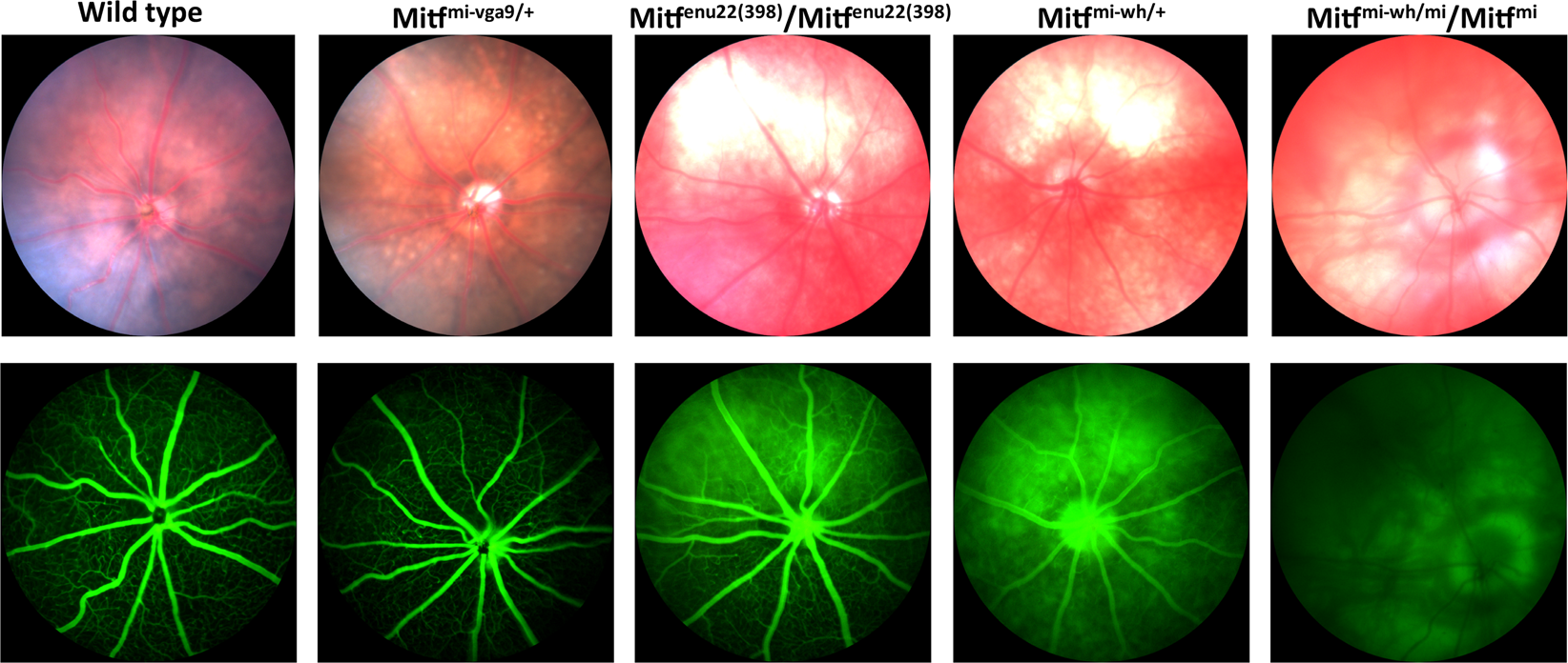
In their paper, “The microphthalmia-associated transcription factor (Mitf) gene and its role in regulating eye function,” García-Llorca et al use the Phoenix MICRON® IV to examine the outer eye appearance, retinal pigmentation, and retinal vasculature through fluorescein angiography to study several different mouse mutants. Combined with electroretinography and histology, the fundus images tell a story of how subtle genetic differences can result in large phenotypic differences.
Retinal degeneration causes visual impairment and blindness and can be caused by mutations in retinal cells including the photoreceptors and retinal pigment epithelium (RPE). The Mitf gene is involved in RPE differentiation and proliferation and mutations in the gene can lead to coat pigmentation abnormalities and inner ear and eye defects. Mutations are associated with the human diseases Waardenburg and Tietz syndrome and as well as small eye defects. García-Llorca et al study four Mitf mutant mice strains with normal eye size to examine the more subtle effects that an RPE gene abnormality might cause.
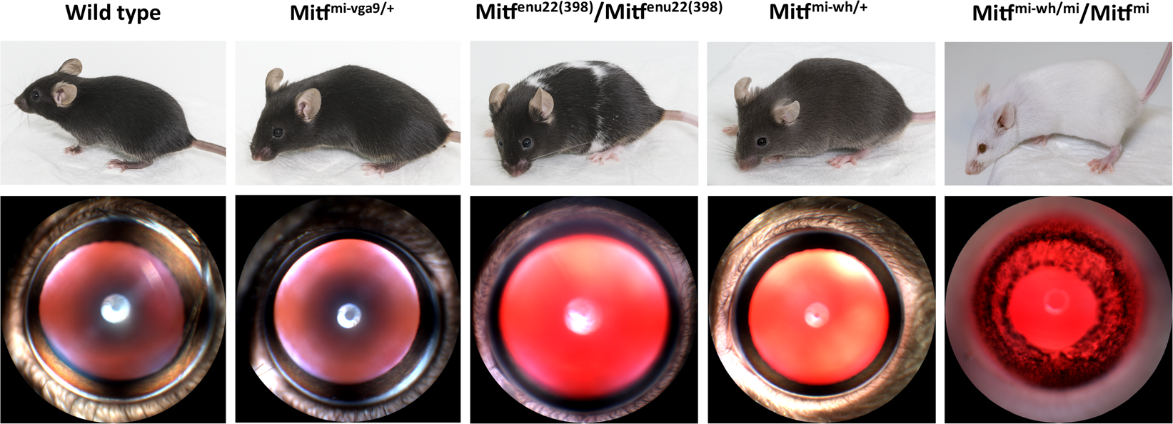
The four strains, with coat pigmentation and outer eye appearance shown in Figure 1, are: heterozygous Mitfmi–vga9/+, homozygous Mitfmi–enu22(398), heterozygous Mitf–Mi–Wh/+, and MitfMi–Wh/Mitfmi. The heterozygous Mitfmi–vga9/+ and homozygous Mitfmi–enu22(398) had normal eye size, normal ERG responses, and mostly normal histology. However, the Phoenix MICRON® IV fundus images revealed that heterozygous Mitfmi–vga9/+ had discrete yellow lesions with normal vasculature while homozygous Mitfmi–enu22(398) had large unpigmented lesions with irregular borders and hyperfluorescent areas in fluorescein angiography, though normal capillaries (Fig 2). The other two mutants had much more dysfunction: no ERG responses with widespread retinal degeneration. The Phoenix MICRON® IV images showed that the heterozygous Mitf–Mi–Wh/+ had hypopigmented retinas with large lack of pigmentation and reduced capillaries while the MitfMi–Wh/Mitfmi had eye dilation problems and a widespread lack of pigmentation in the retina (Fig 2).
As the authors write, “This study provides more evidence that a functional RPE is important for normal photoreceptor function,” as Mitf is RPE-specific and not expressed in the neuroretina. The RPE is essential for normal retinal structure and function and studies such as these examining the various genetic factors affecting the RPE clarify the role of the RPE in normal and dysfunctional eyes.
García-Llorca, A., Aspelund, S. G., Ogmundsdottir, M. H., Steingrimsson, E., & Eysteinsson, T. (2019). The microphthalmia-associated transcription factor ( Mitf ) gene and its role in regulating eye function. Scientific Reports,9(1), 1–12.


