Glaucoma, a leading cause of irreversible vision loss, is characterized by progressive damage to retinal ganglion cells (RGCs) and the optic nerve, often associated with increased intraocular pressure (IOP). While previous studies have implicated Tau protein expression and phosphorylation changes in other neurodegenerative diseases such as Alzheimer’s disease, Parkinson’s disease and glaucoma, the causative role […]
08.04
2024
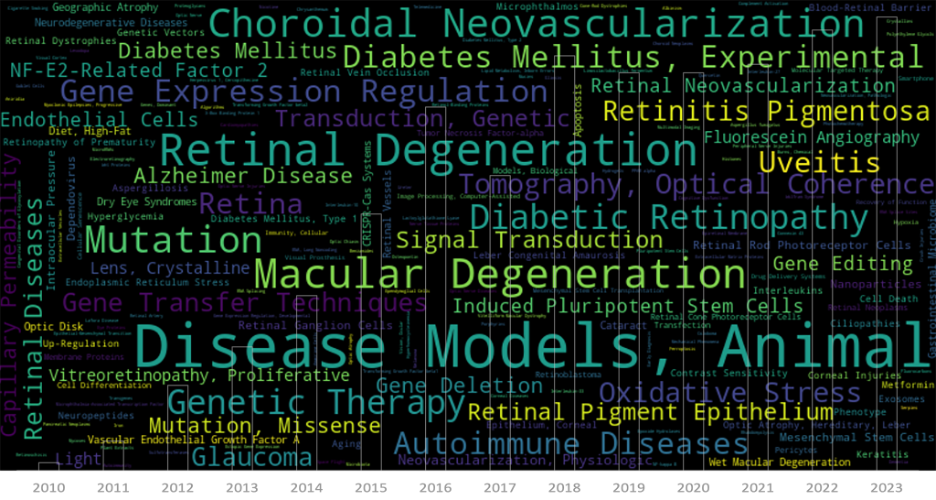
MICRON Imaging Systems: Powering 900 Scientific Publications
The ability to see the world around us is a gift that’s easy to take for granted, until it’s threatened by the onset of a devastating eye disease. For those facing the reality of vision loss, ophthalmic research represents a beacon of hope, offering the promise of new treatments and therapies to preserve or restore […]
15.02
2019
Corneal Thickness Analysis using OCT
Corneal images taken with the Phoenix Micron IV OCT used for thickness analysis King et al, a consortium of researchers at a range of institutions, recently used the Phoenix Micron IV OCT to examine corneal thickness in their article, “Genomic locus modulating corneal thickness in the mouse identifies POU6F2 as a potential risk of developing […]
06.12
2017
Retinal Ganglion Cell Survival During Chronic and Acute Injury
Using the Phoenix retinal imaging and functional measurement to study retinal ganglion cell survival during chronic (glaucoma) and acute (optic nerve crush) injury
Liu et al studied the survival and dysfunction of retinal ganglion cells (RGC) during chronic (glaucoma) and acute (optic nerve crush) injury in a series of comprehensive and elegant articles published from 2015 to 2017. The researchers used the Phoenix ERG and Micron IV provide a complete picture of RGC disruption.
15.08
2017
Studying Glaucoma with Micron IV Fluorescent RGC Imaging
Dendrites may be retracted in several diseases as glaucoma. Studying morphology of dendritic arbors may give us an idea about functional deficits in those diseases. The Di Polo lab at the University of Montreal researches glaucoma using the Micron IV rodent retinal imaging camera and OCT module. Their scientists captured stunning fluorescent images with the Micron IV of mice genetically modified to produce yellow fluorescent protein (YFP)-taged retinal ganglion cells (RGC). the brightest RGC are visible in the bright field image along with blood vessels, optic nerve, and the retinal surface.


