Corneal images taken with the Phoenix Micron IV OCT used for thickness analysis
King et al, a consortium of researchers at a range of institutions, recently used the Phoenix Micron IV OCT to examine corneal thickness in their article, “Genomic locus modulating corneal thickness in the mouse identifies POU6F2 as a potential risk of developing glaucoma.” Thinner central corneal thickness, which is hereditary, is associated with an increased risk of developing primary open angle glaucoma. While many genes are linked to central corneal thickness, they only explain 8% of variability. Using inbred mouse strains, King et al provide compelling evidence for linking the gene Pou6f2 to central corneal thickness, potentially affecting glaucoma development, and even retinal ganglion cell loss during glaucoma.
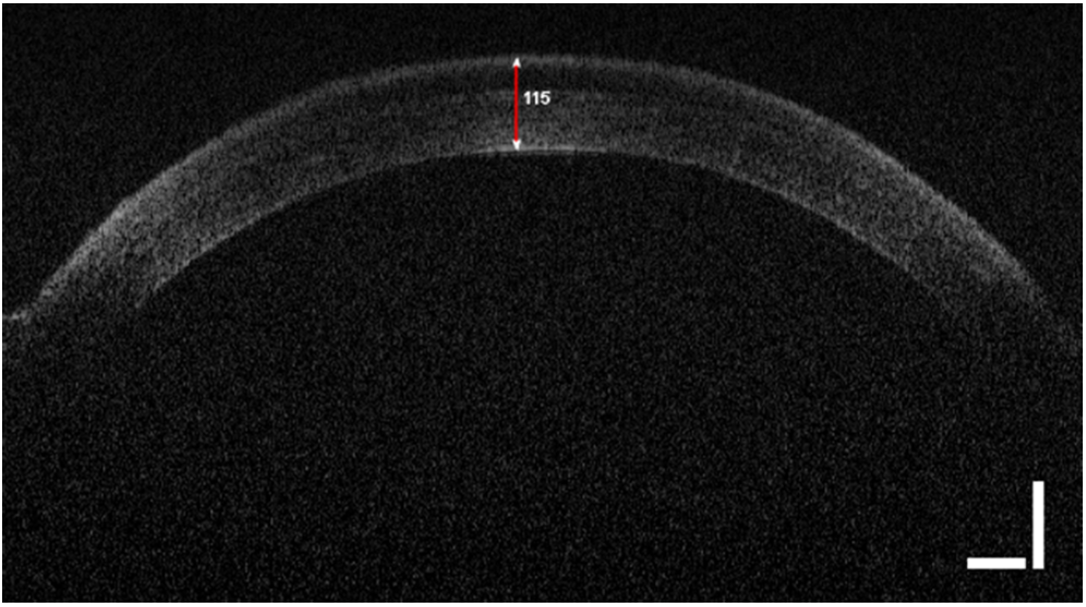
BXI mice are a range of highly inbred substrains produced from the parent strains of C57BL/6J and DBA2/J and whose genomes have been fully mapped. King et al tested a total of 818 mice across 61 substrains of BXD. The Phoenix Micron IV OCT produced clear, crisp images of the cornea (Fig. 1). The included Insight semi-automated segmentation software allows for quantitative retinal and corneal segmentation. Using two OCT systems, including the Phoenix Micron IV OCT, the team found an average central corneal thickness of 100 µm across the BXD strains; the thinnest as 85 µm and the thickest was 125 µm (Fig. 2).
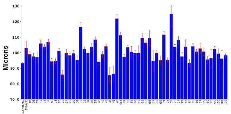
The BXD parent strains’ average thickness is 93 µm for C57BL/6J and 103 µm for DBA. The standard deviation of the central corneal thickness for each of the BXD substrains is small, indicating a strong hereditary component. Using genetic mapping tools, the researchers determined that Pou6f2 may be a candidate for modulating central corneal thickness and thus changing risk for glaucoma and, intriguingly, retinal ganglion cell loss during glaucoma. The gene appears to be preserved in humans as well so may be a potential therapeutic target.
While King et al used the retinal lens to image the cornea, Phoenix has a new anterior segment lens which produces detailed images of the cornea, lens, iris, and even Schlemm’s canal.
Reference: King R, Struebing FL, Li Y, et al. Genomic locus modulating corneal thickness in the mouse identifies POU6F2 as a potential risk of developing glaucoma. PLoS Genet. 2018;14(1):e1007145. Published 2018 Jan 25. doi:10.1371/journal.pgen.1007145