The ability to see the world around us is a gift that’s easy to take for granted, until it’s threatened by the onset of a devastating eye disease. For those facing the reality of vision loss, ophthalmic research represents a beacon of hope, offering the promise of new treatments and therapies to preserve or restore sight.
In a significant milestone, we are excited to share that images and data from MICRON® imaging systems have been included in over 900 scientific research papers and publications. From cutting-edge genetic therapies to novel disease models and documentation of cellular processes, I am proud that our imaging solutions have supported researchers as they endeavor to unlock groundbreaking insights and drive innovation in the field of ophthalmology and neurology.
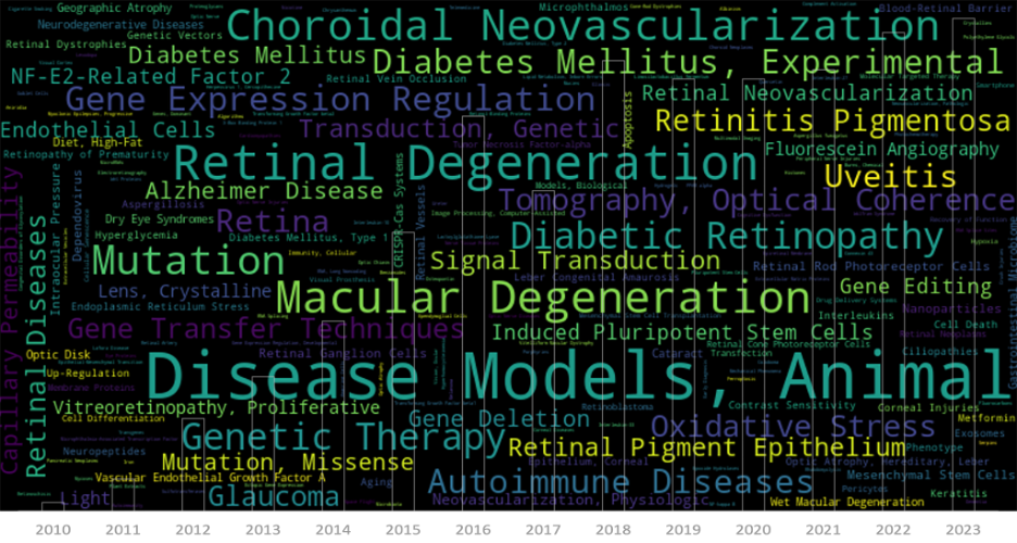
Used to image the eyes of small animals from zebrafish and hummingbirds to rodents and small rabbits, the breadth of pre-clinical studies encompassing MICRON imaging systems is truly remarkable. Researchers have leveraged our technologies to explore animal and disease models for various ophthalmic conditions, including retinal degeneration, macular degeneration, retinal vein occlusion, glaucoma, and retinitis pigmentosa. Our systems have been used to explore how neurologic disorders like Alzheimer’s Disease are expressed in the retina as well as to create novel animal models such as laser-induced geographic atrophy in mice. These investigations have shed light on the underlying mechanisms, diagnostic techniques, and potential therapeutic approaches for these debilitating and sight-threatening diseases.
MICRON imaging solutions have played a role in genetic studies and therapies, enabling researchers to delve into the intricate world of gene expression regulation, mutation analysis, and the effects of gene editing techniques like CRISPR-Cas systems. Not only that, spectacular innovations have been explored using the MICRON in other creative ways such as directly imaging the brain as well as training artificial intelligence (AI) networks to identify disease biomarkers. These advancements hold immense promise for developing targeted treatments and personalizing medicine for ophthalmic disorders.
Cellular and molecular processes in the eye have also been meticulously studied using MICRON imaging systems. Researchers have explored various aspects, such as oxidative stress, signal transduction, neovascularization, and cellular senescence, to unravel the complex mechanisms underlying eye health and disease progression.
As we celebrate this milestone, we are humbled to provide instruments that help our customers in their research journeys. And, I want to reaffirm that in many ways we’re just getting started. Last year we launched the all-new MICRON 5 camera, new lens technology, and MICRON Software Suite, our redesigned image and data management platform. We’ll continue this commitment to innovation as we push the boundaries of eye and eye-brain imaging and data capture, while continuing to provide world-class support to our research customers around the world.
Here’s to supporting the next 900 papers!