In their article, “Effect of MMP-9 gene knockout on retinal vascular form and function,” George et al study the effect of knocking out a matrix protein in a mouse model of retinitis pigmentosa using the Phoenix MICRON® platform including OCT, and Ganzfeld ERG. The combination of the Phoenix MICRON® fundus images, OCT revealing the layers, and Ganzfeld ERG measuring the function gives a complete examination into these mice.
Retinal degeneration is an increasing issue leading to photoreception malfunction and visual impairment. Rd1 mice are a model of retinitis pigmentosa in which the rod cells degenerate and disappear by 3 weeks of age. Matrix metalloproteinase-9 is an extracellular matrix protein, increased levels of which lead to neuroretinal remodeling and significant blood vessel dysfunction. Deceased levels slow down the neuroretinal degenerative process and may help preserve retinal physiology. George et al knocked out MMP-9 on a FVB/NJ background which are monozygous for a faulty rd1 allele and are blind. They studies three mouse strains: C57Bl/6J as control, the retinitis pigmentosa model FVB/NJ, and the MMP-9 KO mice which were the FVB/NJ mice lacking MMP-9.
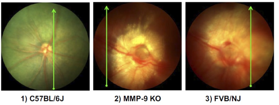
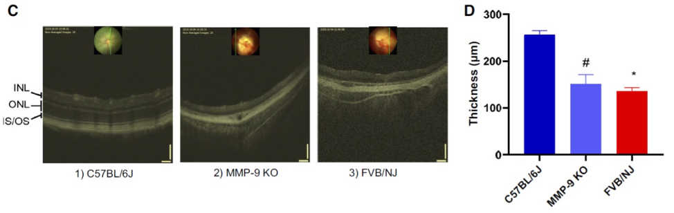
The Phoenix MICRON® fundus images showed large differences among the mouse strains. With detailed images of the vasculature, George et al concluded that the FVP/NJ mice had altered retinal vasculature while the MMP-9 KO had less disruption of vasculature and some vessels preservation (Fig 1). The Phoenix MICRON® OCT delivered clear images of the retinal layers, showing less disruption of layers in MMP-9 KO mouse compared to the extremely disrupted layers and vasculature of FVP/NJ (Fig 2C). The semi-automated segmentation program Insight 2D, exclusively available with the Phoenix MICRON® OCT, revealed that C57Bl/6J have the thickest retinas followed by the MMP-9 KO and then the FVP/NJ mice (Fig 2D). Finally, George et al used the Phoenix Ganzfeld ERG to measure the function of the retinas and found that there was no response in the FVP/NJ mice and a very small response in the MMP-9 KO mice.
Using this complete picture of the retina structure and function, George et al suggest that MMP-9 may be an excellent therapeutic target for retinal degeneration diseases.
George, A. K., Homme, R. P., Majumder, A., Tyagi, S. C., & Singh, M. (2019). Effect of MMP-9 gene knockout on retinal vascular form and function. Physiological Genomics, 51(12), 613–622.