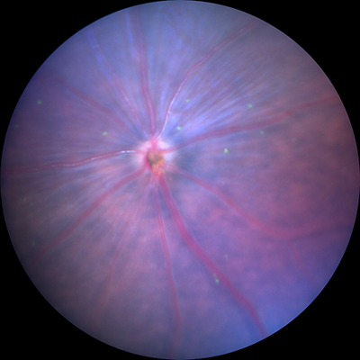
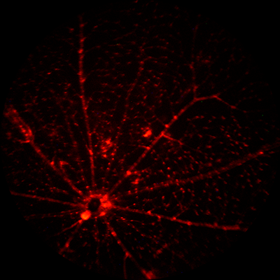
The Di Polo lab at the University of Montreal researches glaucoma using the Micron IV rodent retinal imaging camera and OCT module. Their scientists captured stunning fluorescent images with the Micron IV of mice genetically modified to produce yellow fluorescent protein (YFP)-tagged retinal ganglion cells (RGC). the brightest RGC are visible in the bright field image along with blood vessels, optic nerve, and the retinal surface. In the fluorescent image, the RGC are highlighted and their dendritic processes, appearing as thin lines emanating from the central cell body, are clearly visible. Scientists also examined mice with red fluorescent protein (RFP)-tagged mural cells with the Micron IV; the mural cells clearly highlighted surrounding the vasculature in the fluorescent image.
The Di Polo lab studies the effect of glaucoma on retinal ganglion cells, working toward a treatment to prevent neuronal cell death. The Micron IV will aid in their glaucoma research by allowing them to observe RGC over time as the disease progresses. Using geographic markers on the retina, the same RGC can be monitored longitudinally, especially with simple switching of filters from bright field to fluorescent while the mouse stays in the same position. They will also use the OCT to observe the irido-corneal angle over time.


