Retinal vein occlusion is the second most common retinal vascular disorder leading to loss of vision in developed countries (after diabetic retinopathy). A lack of treatment options is partially caused by a lack of effective animal models; most rodent models do not show the essential symptom of cystoid edema and often heal quickly. In their […]
28.10
2020
A year-long longitudinal pattern dystrophy fundus study with the Phoenix MICRON® IV imaging platform
In their 2019 paper, “Novel molecular mechanisms for Prph2‐associated pattern dystrophy,” Chakraborty et al use the Phoenix MICRON® IV retinal imaging platform to longitudinally study the effect of a very specific mutation affecting the Peripherin 2 protein. Peripherin 2 is a protein in rods and cones which, if mutated, can lead to retinitis pigmentosa, cone-rod […]
17.01
2019
Characterizing a mutant rat strain with the Phoenix Micron OCT and Ganzfeld ERG
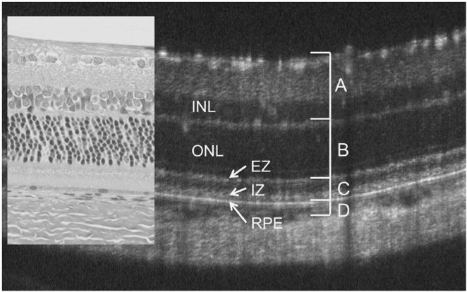
Monai et al characterized the longitudinal retinal degeneration of a rat model of retinitis pigmentosa using the Phoenix Micron OCT to examine retinal layers in live rats and the full field Ganzfeld ERG to test function. The rats have one of the mutations, P23H, that cause retinitis pigmentosa in humans, and are specifically a very […]


