Dr. Rafael Ufret-Vincenty’s lab at University of Texas Southwestern Medical Center has developed a novel model for light damage using the Micron IV rodent retinal imaging camera. This quick and consistent light damage model leads to fundus abnormalities and retinal thinning as measured by the Micron image-guided OCT and semi-automated layer analysis tool, Insight. In two elegant articles, the researchers provided proof of concept in pigmented mice, which are a better model for human eye light damage than overly sensitive albino mice, which demonstrated bleached fundus and outer retinal layer thinning.
06.12
2017
Retinal Ganglion Cell Survival During Chronic and Acute Injury
Using the Phoenix retinal imaging and functional measurement to study retinal ganglion cell survival during chronic (glaucoma) and acute (optic nerve crush) injury
Liu et al studied the survival and dysfunction of retinal ganglion cells (RGC) during chronic (glaucoma) and acute (optic nerve crush) injury in a series of comprehensive and elegant articles published from 2015 to 2017. The researchers used the Phoenix ERG and Micron IV provide a complete picture of RGC disruption.
02.11
2017
Micron Reveals Decreased Retinal Ganglion Cell Arborization in a Mouse Model of Retinal Ischemia
Researchers Dailey et al, in the Mitton Lab at Oakland University used the Micron retinal imaging camera to examine retinal ganglion cell (RGC) survival in a mouse model of retinal ischemia. Oxygen-induced retinopathy (OIR) in mice recapitulates critical factors of the human diseases retinopathy of prematurity and diabetic retinopathy. Mice pups were raised in hyperoxegenated air (75% oxygen) for five days and then returned to room air (20% oxygen), which lead to pathological changes in the vascular and neural growth.
15.08
2017
Studying Glaucoma with Micron IV Fluorescent RGC Imaging
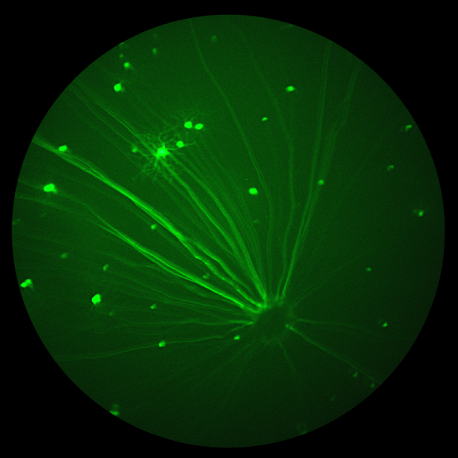
Dendrites may be retracted in several diseases as glaucoma. Studying morphology of dendritic arbors may give us an idea about functional deficits in those diseases. The Di Polo lab at the University of Montreal researches glaucoma using the Micron IV rodent retinal imaging camera and OCT module. Their scientists captured stunning fluorescent images with the Micron IV of mice genetically modified to produce yellow fluorescent protein (YFP)-taged retinal ganglion cells (RGC). the brightest RGC are visible in the bright field image along with blood vessels, optic nerve, and the retinal surface.
07.03
2017
Micron Retinal Imaging System Provides Ground Breaking Research on Parkinson’s Disease
Phoenix Research Labs is pleased to announce ground breaking research on Parkinson’s Disease by Price et al using the Micron Retinal Imaging System. Abnormally high concentration of the protein a-synuclein in the brain is linked with the physical and mental deficits caused by Parkinson’s Disease {PD) and Dementia with Lewy Bodies (DLP).
29.11
2016
Detection of Infectious Diseases in the Retina of Mice
A poster presentation by Emily Gordon, et al., from the National Institute of Allergy and Infectious Diseases revealed the observation of cerebral malaria in the mouse retina as imaged by the Phoenix Research Labs retinal camera the Micron IV.
According to the authors “Microcirculation in the retinal vasculature provides a window to image dynamic changes taking place in the central nervous system during CM disease progression.


