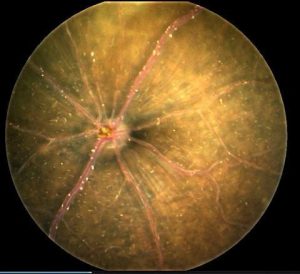
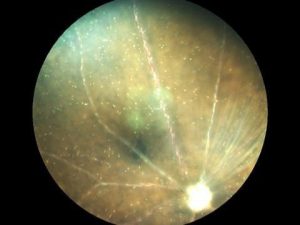
A poster presentation by Emily Gordon, et al., from the National Institute of Allergy and Infectious Diseases revealed the observation of cerebral malaria in the mouse retina as imaged by the Micron retinal camera.
According to the authors “Microcirculation in the retinal vasculature provides a window to image dynamic changes taking place in the central nervous system during CM disease progression.” In particular, the authors state that “brightfield fundoscopy reveals flowing hyper-reflective clumps that are confined to the retinal vasculature” and that these are only present in infected animals.
Such observations open up the perspective that infectious diseases can be monitored by retinal microscopy.
This kind of monitoring was also reflected in an earlier release of observations of Alzheimers and Parkinson’s disease through retinal imaging. (See Alzheimer’s and Parkinson’s research).
Phoenix, as does authors of this presentation, sees the potential to monitor many diseases through retinal observations.


