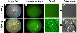
Phoenix Research Labs is pleased to announce ground breaking research on Parkinson’s Disease by Price et al using the Micron Retinal Imaging System. Abnormally high concentration of the protein a-synuclein in the brain is linked with the physical and mental deficits caused by Parkinson’s Disease {PD) and Dementia with Lewy Bodies (DLP). A mouse model overexpressing GFP – tagged a-synuclein, a-syn::GFP (Fig 1, above), recapitulates many of the clinically relevant physical issues seen in PD and DLP. The Micron clearly shows the a-synuclein punctae in the retina, allowing a glimpse of the brain through the eyes.
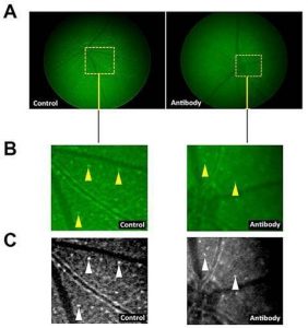
Researchers Price et al examined the retinas of a-syn::GFP mice using the Micron Retinal Imaging Camera and found a significant increase in the number of a-synuclein puntae as the mice aged. Additionally, an immunotherapy against a-synuclein reduced the number of punctae in the retina, revealing a potential clinical therapy for those suffering from disorders associated with overabundance of a-synuclein.
The Micron Retinal Imaging System provided crisp brightfield and GFP images of a-synuclein and allowed longitudinal studies of potential clinical therapies (Fig4, right). Imaging retinas of diseased mice using the Micron III is a powerful way to study the eye-brain connection.