Researchers Dailey et al, in the Mitton Lab at Oakland University used the Micron retinal imaging camera to examine retinal ganglion cell (RGC) survival in a mouse model of retinal ischemia. Oxygen-induced retinopathy (OIR) in mice recapitulates critical factors of the human diseases retinopathy of prematurity and diabetic retinopathy. Mice pups were raised in hyperoxegenated air (75% oxygen) for five days and then returned to room air (20% oxygen), which lead to pathological changes in the vascular and neural growth
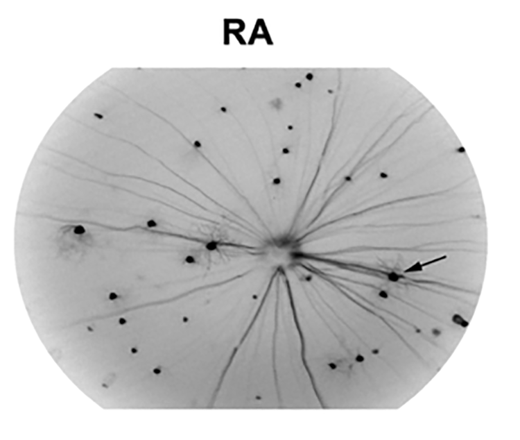 The Micron retinal imaging camera shows stunning detail of RGC dendritic arbors in normal (RA room air) mice, allowing for longitudinal studies
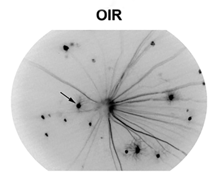 The Micron retinal imaging camera shows stunning detail of diseased (OIR oxygen-induced retinopathy) mice, allowing for longitudinal studies
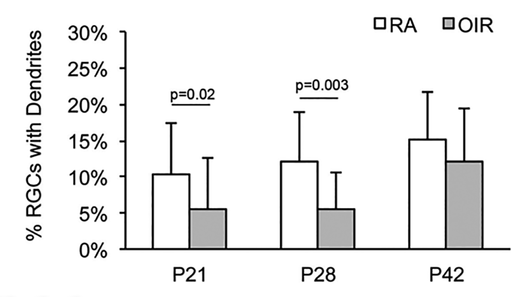 Diseased (OIR oxygen-induced retinopathy) mice had significantly fewer RGC with dendritic arborization at P21 and P28 than normal (RA room air) mice.
Dailey et al examined the structure and histology of the retina, particularly the RGC, after oxygen-induced retinopathy and with treatment with Norrin, a neuroprotective Wnt-signaling ligand. Thy1-YFP mice express YFP in retinal ganglion cells, allowing longitudinal live visualization of RGC. The greyscale images show stunning resolution of the dendritic arbors of RGC in diseased and normal mice (Fig 1). These detailed images revealed that at P21 and P28, there were significantly fewer RGC with dendritic arborization in oxygen-induced retinopathy mice compared to the normal room air mice (Fig 2).
Other findings include disruption and thinning of retinal layers in diseased mice. Histology revealed that Norrin increased RGC survival in diseased mouse eyes.


