In their paper, “Connexin43 Mimetic Peptide Improves Retinal Function and Reduces Inflammation in a Light-Damaged Albino Rat Model,” Guo et al promoted neuroretinal survival in a light damage paradigm by blocking the Connexin43 hemichannels. Using the Phoenix MICRON® OCT to examine the structure of the retina at precise retinal locations as shown by the bright field fundus image, Guo et al demonstrated that the retinal degeneration caused by light damage was halted by treatment. Insight 2D, a semi-automated retinal layer segmentation tool that is exclusively provided with the Phoenix MICRON® OCT to analyze rodent OCT images, was used to precisely quantify the thinning of the damaged and treated retinas. Rounding out the study, electroretinography showed preserved function from the treatment while immunohistochemistry revealed the decrease in inflammation in the retina after treatment.
The choroid provides oxygen and nutrients to the outer retina and is degenerated in diseases such as diabetic retinopathy and age-related macular degeneration. Connexin43 is a ubiquitous gap junction protein with a big role in the inflammatory response. Under normal conditions, Connexin43 hemichannels are closed but under inflammatory stress, the channels open and lead to cell-damaging cascades. Peptide5 is a Connexin43 mimetic peptide that reduces inflammation by blocking Connexin43 hemichannels and has a neuroprotective effect in spinal cord injury, cerebral ischemia, and retinal ischemia-reperfusion.
Guo et al exposed albino rats to 2700 lux fluorescent lamps for 24 hours. Two hours after the start of light exposure, they intravitreally injected Peptide5 or a sham treatment.
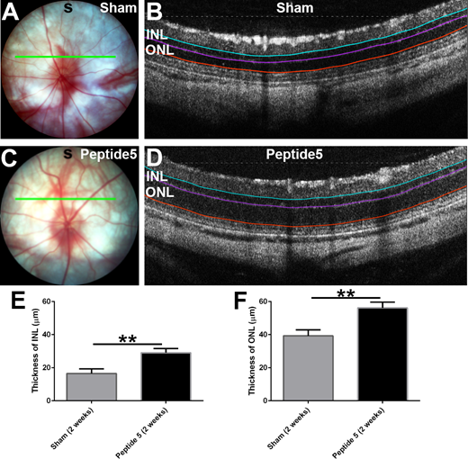
Figure 1. The Phoenix MICRON® OCT live fundus image (A&C) with corresponding OCT image (B&D) under the thin green line. Peptide5 treatment (C&D) preserved retinal structure compared to sham treatment (A&B). The teal, purple, and orange lines on the OCT image are from the exclusive Insight 2D semi-automated segmentation software. E&F: Associated quantitative values of inner nuclear retina thickness (E) and outer nuclear retina thickness (F).
In Figure 1, the Phoenix MICRON® live bright field retinal image (Fig 1 A&C) is shown to the left of the Phoenix MICRON® OCT image (Fig 1 B&D). The thin green line on the fundus is where the OCT scan is taken and can be moved around in real-time. The teal, purple, and orange lines on the OCT image are layers outlined and measured with Insight 2D; the exclusive semi-automated OCT analysis program segments the layers and provides both qualitative (Fig 1 B&D) and quantitative (Fig 1 E&F) data. Guo et al used the Phoenix MICRON® OCT and Insight 2D to discover that two weeks after light damage and treatment, the inner and outer nuclear layers thinned less after Peptide5 treatment (Fig 1 C&D) compared to sham treatment (Fig 1 A&B). Peptide5 thus protected retinal neurons from degeneration.
Rounding out the study with structure, function, and cell-level analysis, Guo et al found that rod and cone electroretinographical responses were decreased after light damage but preserved with Peptide5 treatment. Similarly, inflammatory cells were elevated in the choroid and retina but improved with treatment.
In this study, blocking Connexin43 hemichannels resulted in better functional and structural outcomes in both the short and long term, showing a possible pathway for treatments of diabetic retinopathy, diabetic macula edema, and age-related macular degeneration.
Citation:
Guo, C. X., Mat Nor, M. N., Danesh-Meyer, H. V., Vessey, K. A., Fletcher, E. L., O’Carroll, S. J., Acosta, M. L., & Green, C. R. (2016). Connexin43 Mimetic Peptide Improves Retinal Function and Reduces Inflammation in a Light-Damaged Albino Rat Model. Investigative Ophthalmology & Visual Science, 57(10), 3961–3973.