In their paper “Quantitative Analysis of Retinal Structure and Function in Two Chromosomally Altered Mouse Models of Down Syndrome”, researchers Victorino, Scott-McKean, et al leveraged the multi-modality capabilities of the Phoenix MICRON™ retinal imaging platform, to produce an image-rich research paper looking at the ocular features of Down Syndrome in two mouse models; Ts65Dn and Dp16.
Down Syndrome (DS), or Trisomy 21 is one of the most common genetic abnormalities in humans, occurring in approximately 1 in 700 live births. Physical growth delays, intellectual impairments and distinct facial features are hallmarks of the syndrome. Patients with DS may also present with abnormalities in eye structure and function including refractive errors, corneal ectasia, cataracts and retinovascular anomalies. The question was to understand if ocular features were mimicked in these mouse models
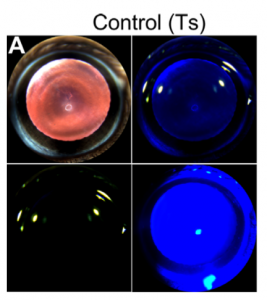
Using the Phoenix MICRON slit lamp, cobalt blue filter and red reflex color photos were captured to document potential signs of cataracts and corneal changes in the mouse models and respective controls. Although 60% of DS patients present with these findings, no significant pathology was noted in the mouse cohorts.
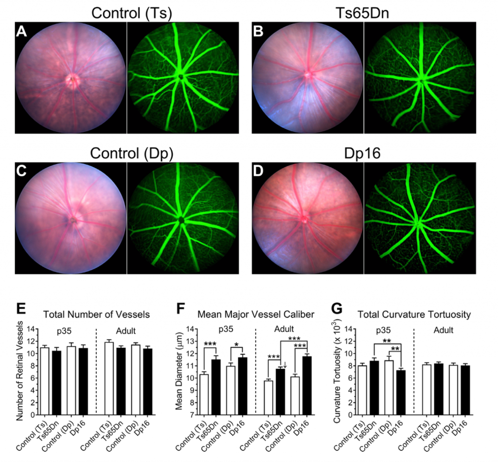
Beautiful Micron IV bright field retinal photos and fluorescein angiography images were foundations in the new identification of enlarged retinal blood vessels. Vascular parameters were quantified including total number of vessels, vessel caliber, tortuosity and fractal dimension.
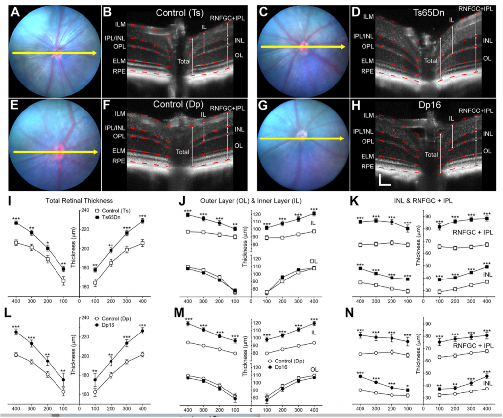
The MICRON OCT attachment provided image-guided optical sections of retinal thicknesses, measured using Phoenix InSight software, confirming findings of increased inner retinal thickness in the two DS mouse models.
Notably, the MICRON Ganzfeld ERG enabled the first characterization of retinal function in Dp16 mice. Additionally, Victorini et al were able to test and illustrate the effects of different anesthetic agents on the properties of ERGs to further elucidate discrepancies found between previous work and another study.
The experience described in this paper provides beautiful visuals as evidence of the similarities and differences in the mouse DS models and humans, and the suggestion to consider the influence of anesthesia agents on ERG outcomes.
Citation: Victorino DB, Scott-McKean JJ, Johnson MW, Costa ACS. Quantitative analysis of retinal structure and function in two chromosomally altered mouse models of Down syndrome. Invest Ophthalmol Vis Sci. 2020;61(5):25. https://doi.org/10.1167/iovs.61.5.25


