In a recent well written, compelling article published in Nature Communications, “Endothelial activation of caspase-9 promotes neurovascular injury in retinal vein occlusion,” Avrutsky et al show that caspase-9 inhibition is a promising treatment for retinal vein occlusion. Retinal vein occlusion models hypoxic-ischemic neurovascular damage and is the second leading cause of blindness in working-age adults. Currently, treatment consists of anti-VEGF drugs which lead to inconsistent physiological and functional recovery. As caspase-9 expression is increased with hypoxic-ischemia, Avrutsky et al used caspase-9 inhibiting eyedrops, Pen1-XBir3, after retinal vein occlusion in mice.
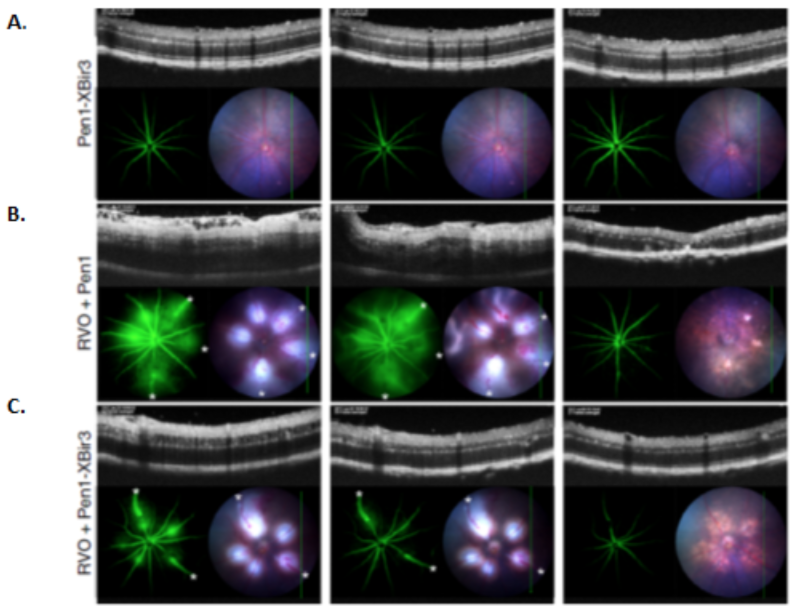
The Phoenix MICRON® image-guided laser was used to create retinal vein occlusion by targeting and ablating the major retinal veins of the mouse eyes (Fig 1B). Directly after and one day post-the laser injury, mice received the caspase-9 inhibitor Pen1-XBir3 or a sham eyedrop treatment. The Phoenix MICRON® OCT revealed the subsequent retinal detachment, swelling, and disorganization of retinal inner layers after retinal vein occlusion (Fig 1B) as well as the presence of hyper-reflective foci—though there is debate about what the foci are, they are a measure of retinal inflammation and the degree of vision recovery after retinal vein occlusion in humans. Insight 2D, a semi-automated segmentation program included with the Phoenix MICRON® OCT, was used to measure the thickness of the retinal layers before and after retinal vein occlusion. Pen1-XBir3 treatment reduced the retinal swelling, accumulation of retinal fluid, and retinal detachment, preserving photoreceptor health (Fig 1C).
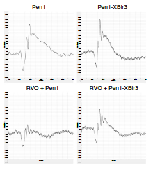
One, two and eight days after injury, the mice were injected with fluorescein and examined with the Phoenix MICRON to measure retinal blood vessel leakage, which Pen1-XBir3 significantly reduced (Fig 1B and 1C). To assess functional damage and recovery with treatment, the Phoenix MICRON® focal ERG, with which the location of a focal white light stimulus is chosen under dark-red illumination to preserve dark adaptation, revealed that while retinal vein occlusion decreases b-wave amplitude (and to a lesser extent, a-wave amplitude), the Pen1-XBir3 treatment recovers the b-wave function (Fig 2).
Using most of the suite of Phoenix MICRON® products, Avrutsky et al demonstrated a powerful treatment for retinal vein occlusion that preserves the structure and function of the retina after intense vascular damage.
Avrutsky, M. I., Ortiz, C. C., Johnson, K. V., Potenski, A. M., Chen, C. W., Lawson, J. M., White, A. J., Yuen, S. K., Morales, F. N., Canepa, E., Snipas, S., Salvesen, G. S., Jean, Y. Y., & Troy, C. M. (2020). Endothelial activation of caspase-9 promotes neurovascular injury in retinal vein occlusion. Nature Communications, 11(1), 3173


