Researchers Lin et al at Taipei Medical University shared an alarming finding that blue light, similar to that emitted by smart phones, can lead to retinal disruption in rats. They used the Phoenix-Micron’s Phoenix MICRON® fundus camera and the image-guided OCT to demonstrate blood vessel leakage and retinal thinning after intermittent blue light exposure.
Lin et al first showed reduced viability of the retinal pigment epithelium (RPE) in cell culture when the cells were exposed to blue light. Selected apoptotic pathways were activated, leading to cell damage and death. They then exposed Brown Norway rats to 460 nm blue light at a 150 lux level in four groups: 0 hours, 0.5 hours, 1 hour, and 3 hours per day. Imaging timepoints were at 0 days, 14 days, and 28 days.
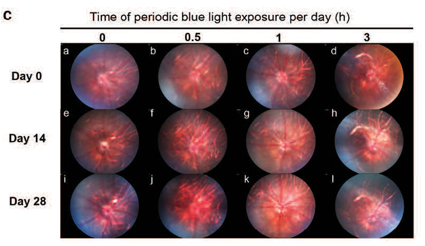
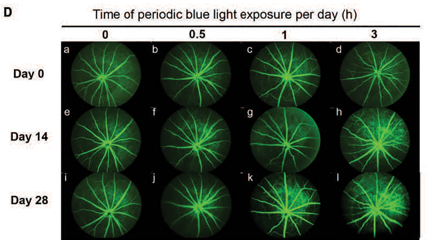
Fluorescein angiography taken with the Phoenix MICRON® fundus camera, which allows crystal clear, live video and still imaging of rodent retinas, shows leakage in the 1 hour of blue light per day rat group at day 28 (Figure 1k). Logically, the 3 hours per day rat group had earlier evidence of leakage at day 14 and 28 (Figure 1h, 1l). Brightfield fundus imaging of the same rats shows some damage of the 3 hour per day rats at day 28 (Figure 2l).
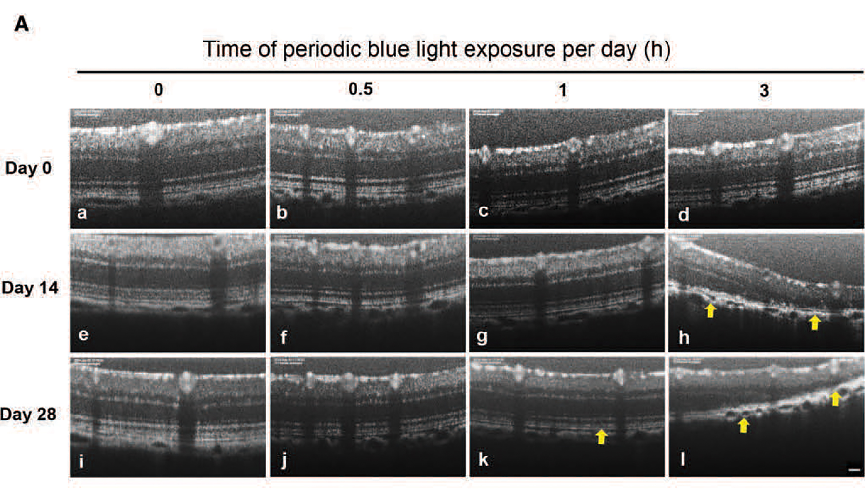
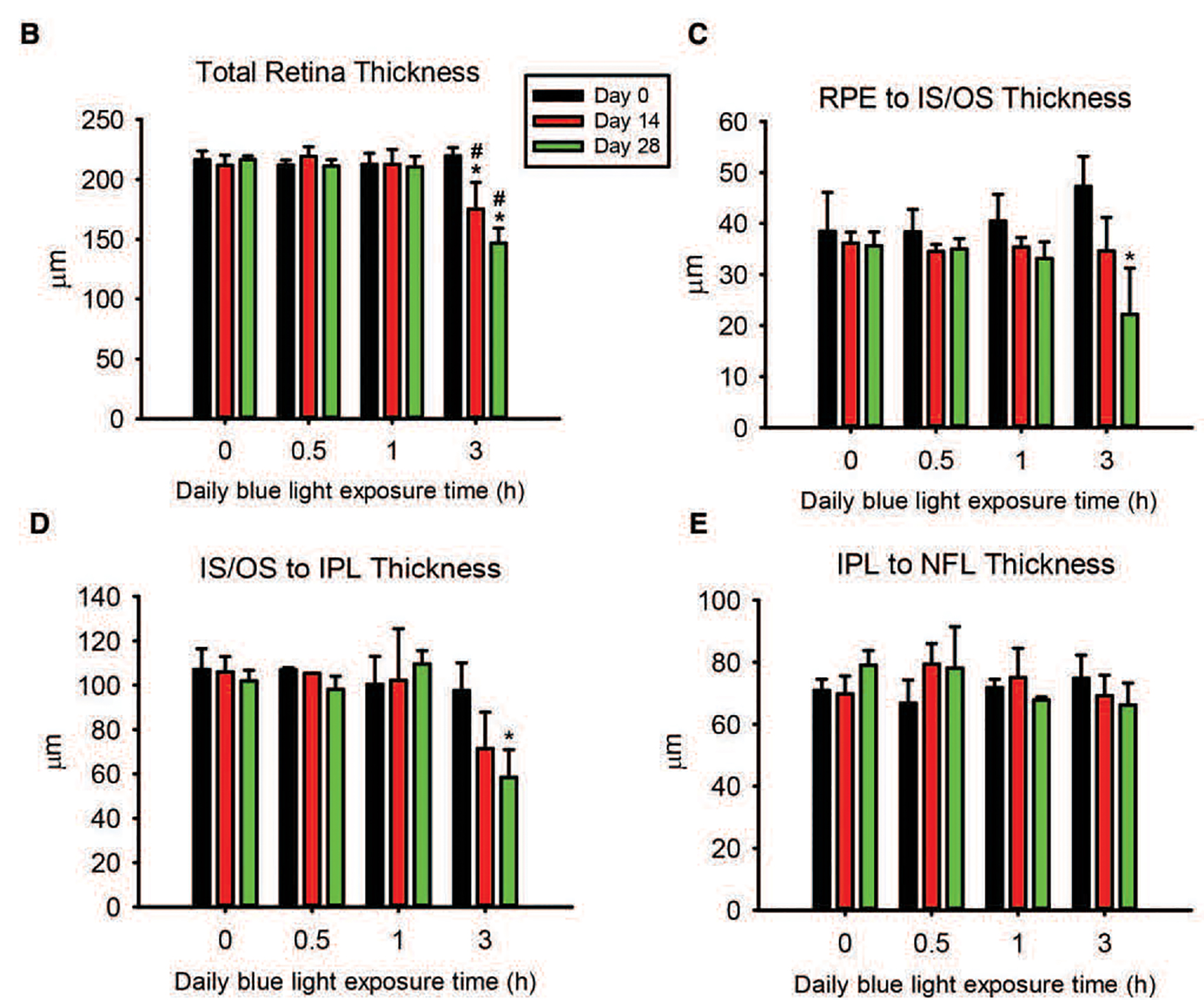
The OCT images show the most dramatic changes. The Phoenix MICRON® OCT provides high resolution, live OCT imaging with a corresponding fundus image for precise location-specific retinal OCT examination. In the 3 hours of blue light exposure per day rats, the OCT reveals retinal layer and choroid disruption, interneuron layer loss, dim rods and cones, and overall retinal thinning (Figure 3h, 3l). The 1 hour of blue light per day rats showed similar disruption, but at a later time point (Figure 3k). Quantification of the layer thicknesses shown in Figure 4.
ERG recordings showed decreased a- and b-waves in all groups at 28 days.
Taken together, these data indicate that exposure to blue light similar to that emitted by smart phone screens may lead to retinal degeneration and even, Lin et al postulate, dry-age related macular degeneration.


