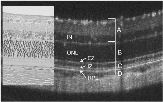
In the July edition of Nanomedicine journal, Chemla et al demonstrate a fascinating and novel way to label and track individual photoreceptor precursor cells migrating within the retina with fluorescence and gold nanoparticle tagging using the Phoenix MICRON® and OCT. Many retinal diseases such as age-related macular degeneration and retinitis pigmentosa are characterized by photoreceptor
26.08
2019
The Secret to a Perfect Fundus Image
Hint: It Starts with the Animal Angle During our hands-on training, we review animal positioning until every user is completely comfortable. But like most other lab equipment task, it does take some practice. Aligning the animal just right can be the difference between a great and a good fundus image. We’ve put together a
14.05
2019
ARVO 2019: Frogs, Glue and CNV
Another ARVO passed by in a blur of research, scientific discussions, and seeing science friends. If you came by our booth, thank you for swinging by to chat with the Phoenix team. If you didn’t get a chance, please let me know if you have any questions about the Micron system and look for our
27.03
2019
Phoenix MICRON® CNV System used to test a novel treatment for age-related macular degeneration
In their paper, “Suppression of Choroidal Neovascularization by AAV-Based Dual-Acting Antiangiogenic Gene Therapy,” Askou et al develop an adeno-associated virus (AAV) treatment for age-related macular degeneration. Beautiful fluorescent fundoscopy performed with the Phoenix MICRON® validated the success of the subretinal AAV injection, while precise choroidal neovascularization (CNV) induced by Phoenix laser burns confirmed that the
15.02
2019
Corneal Thickness Analysis using OCT
Corneal images taken with the Phoenix Micron IV OCT used for thickness analysis King et al, a consortium of researchers at a range of institutions, recently used the Phoenix Micron IV OCT to examine corneal thickness in their article, “Genomic locus modulating corneal thickness in the mouse identifies POU6F2 as a potential risk of developing
17.01
2019
Characterizing a mutant rat strain with the Phoenix Micron OCT and Ganzfeld ERG
Monai et al characterized the longitudinal retinal degeneration of a rat model of retinitis pigmentosa using the Phoenix Micron OCT to examine retinal layers in live rats and the full field Ganzfeld ERG to test function. The rats have one of the mutations, P23H, that cause retinitis pigmentosa in humans, and are specifically a very
