In the July edition of Nanomedicine journal, Chemla et al demonstrate a fascinating and novel way to label and track individual photoreceptor precursor cells migrating within the retina with fluorescence and gold nanoparticle tagging using the Phoenix MICRON® and OCT. Many retinal diseases such as age-related macular degeneration and retinitis pigmentosa are characterized by photoreceptor layer deterioration with healthy photoreceptor-support layers. Despite the relative health of most of the layers of the retina, the photoreceptor degeneration leads to impaired vision. A promising therapy is transplanting photoreceptor-precursor cells into the retina and being able to track their location and survival is essential. Chemla et al developed a cell-tagging mechanism with gold nanoparticles that are visible with OCT and CT scans.
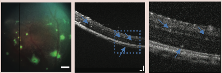
The most common tagging mechanism is with fluorescent proteins, but the fluorescence may not be visible with fundus imaging if the eye has cataracts, the tagged cells are beneath the retinal pigment epithelium, or if the fluorescence is too dim and diffuse. The Phoenix MICRON® produces beautiful images of fluorescently tagged cells in clear eyes as demonstrated in Figures 1 and 2. In an OCT image, fluorescently tagged cells blend into the retina and cannot be definitively identified. Chemla et al elegantly circumvented these issues by tagging their photoreceptor precursor cells with green fluorescent protein (GFP) as well as gold nanoparticles. Since the gold nanoparticles are solid, they are visible in an OCT image which allows for precise and detailed imaging of the cells as they integrate into the retina. They are nontoxic and long lasting; this technique could revolutionize tracking cells in the retina.
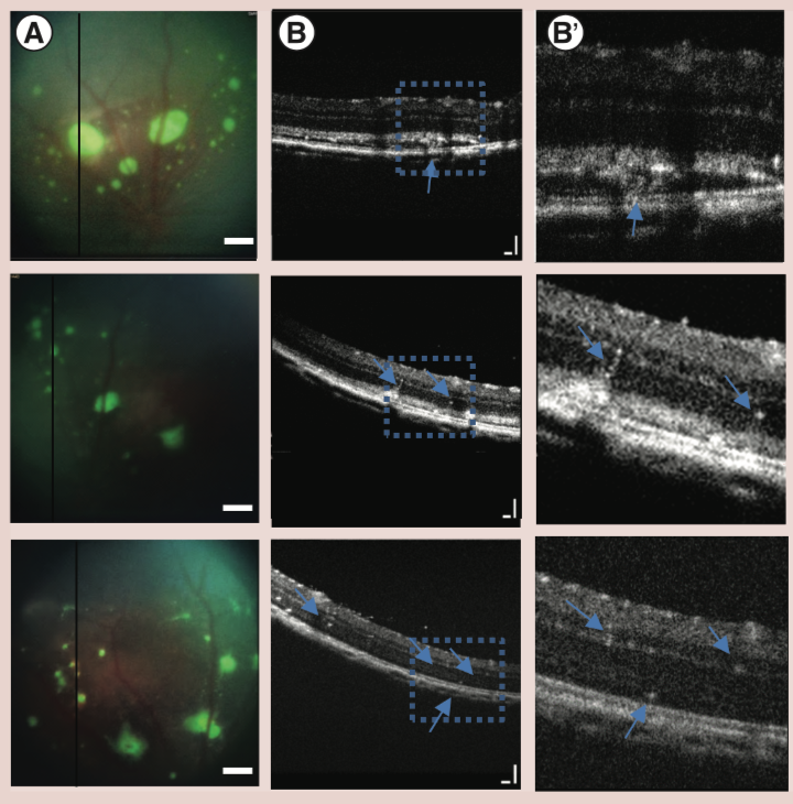
Chemla et al injected the gold nanoparticle-tagged photoreceptor precursor cells subretinally or intravitreally into the eyes of Long-Evans rats and tracked the cells over 30 days. With the subretinal injection, discrete GFP-labeled cell clusters were visible using the Phoenix MICRON® fundus camera throughout the 30 days (Figure 1, column A). Interestingly, the Phoenix MICRON® OCT showed incredible detail of the gold nanoparticle-labeled cells migrating from the injection point to the inner retinal layers (Figure 1, column B and B’). Without the gold nanoparticle labeling, the migrating cells are not visible using OCT. After the intravitreal injection, Phoenix MICRON® fundus photography revealed diffuse GFP signal throughout the vitreous (Figure 2, column A), while the Phoenix MICRON® OCT showed clusters of gold nanoparticle-labeled cells in the vitreous just above the retina (Figure 2, column B and B’).
Chemla et al demonstrated a novel, powerful technique for labeling with gold nanoparticles and tracking cells longitudinally in the retina using the Phoenix MICRON® OCT.
Citation: Chemla Y, Betzer O, Markus A, Farah N, Motiei M, Popovtzer R, Mandel Y. (2019). Gold nanoparticles for multimodal high-resolution imaging of transplanted cells for retinal replacement therapy. Nanomedicine 14(14):1857-1871.


