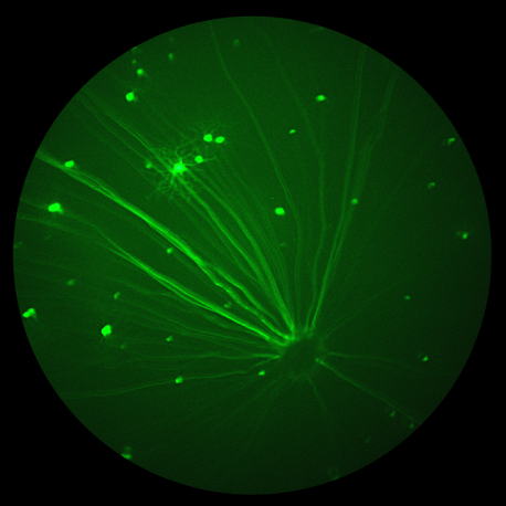
The all-new MICRON® 5 platform delivers advanced performance, repeatability, and imaging workflow efficiency Bend, OR, USA, April 21, 2023 – Phoenix-Micron, Inc. today announced the launch of the all-new Phoenix MICRON® 5 imaging system, the latest breakthrough in small animal ophthalmic imaging technology. With over 450 published studies utilizing MICRON imaging systems, the MICRON platform […]
Phoenix MICRON Research Blog
21.04
2023
Phoenix-Micron, Inc. Announces New, Comprehensive Imaging Software and Redesigned Lens Technology for the Phoenix MICRON® Imaging Platform
Two new innovations, MICRON Software Suite and MICRON LT2 lens technology, streamline the ophthalmic imaging workflow and deliver new data management capabilities. Bend, OR, USA, April 21, 2023– Phoenix-Micron, Inc., a leader in ophthalmic imaging technology, today announced the release of the new MICRON® Software Suite and the MICRON LT2 lens technology. The new […]
08.03
2023
Researchers at the University of Copenhagen demonstrate for the first time that diet induced obesity (DIO) leads to raised intracranial pressure (ICP) and subsequent clinically relevant symptom development.
In the paper “The impact of obesity‑related raised intracranial pressure in rodents” published in Scientific Reports journal, Westgate and collaborators used the Phoenix MICRON® image-guided OCT system, along with the Phoenix MICRON® Insight software, in the measurement of the retinal anatomical changes caused by intracranial hypertension in obese rats. The Problem: Intracranial pressure (ICP) measures the […]
13.10
2022
Where do research ideas come from?
Where do research ideas come from? Do they arrive in a thunderbolt eureka moment, or do they start as an observation or hunch, requiring some time to simmer, brew, incubate and grow? Some may evolve from an accidental or serendipitous discovery during a routine process. Observant operators of the Phoenix MICRON® retinal imaging system may […]
28.02
2022
Analysis of imaging, structure and function using Phoenix MICRON™ modalities expand the understanding of ocular features of Down Syndrome in mouse models
In their paper “Quantitative Analysis of Retinal Structure and Function in Two Chromosomally Altered Mouse Models of Down Syndrome”, researchers Victorino, Scott-McKean, et al leveraged the multi-modality capabilities of the Phoenix MICRON™ retinal imaging platform, to produce an image-rich research paper looking at the ocular features of Down Syndrome in two mouse models; Ts65Dn and […]
19.01
2022
Deep learning provides unbiased evaluation of uveitis severity levels in 1500 Phoenix MICRON® mouse fundus images
In 2020, researchers Jian Sun, Xiaoqin Huang et al, in the Laboratory of Immunology at the National Eye Institute, confirmed in the lab what has been going on in human ophthalmology in recent years, but with a twist. Their deep learning model enabled identification of retinal disease stages with noteworthy accuracy. What marked their work […]


