In their article, “CRYβA3/A1-Crystallin Knockout Develops Nuclear Cataract and Causes Impaired Lysosomal Cargo Clearance and Calpain Activation,” Hegde et al use the Phoenix MICRON® IV Slit Lamp to examine the effect of knocking out a lens structural protein.
α, β and γ crystallins are lens structural proteins that are needed for transparency and refractive power of the lens. Specifically, βΑ3/A1-crystallin is important for lens development and lens structure, but its exact role and method of working is not known. βΑ3/A1-crystallin is present in the lens, retinal astrocytes, retinal pigment epithelium (RPE), and retinal ganglion cells.
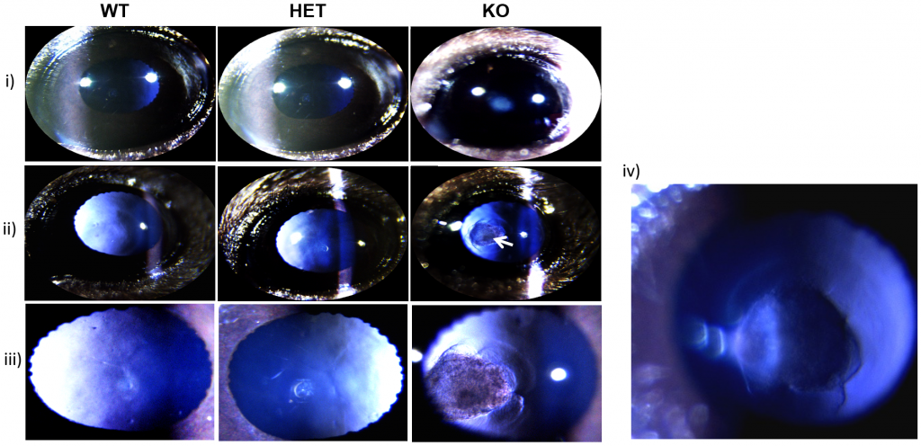
To study the role of βΑ3-crystallin role in the lens, Hedge et al made a βΑ3-knockout mouse. They examined the mice with the Phoenix MICRON® VI Slit Lamp and did extensive immunohistochemical staining and analysis. The Phoenix MICRON® IV Slit Lamp is specifically designed for mice and rats with an adjustable width, length, brightness, and position slit light that clearly illuminates the anterior segment including the lens as used in this study. Although Hedge et al only used the brightfield functionality, the Phoenix MICRON® IV Slit Lamp can also image GFP and fluorescein angiography.
As seen in Figure 1, 100% of βΑ3-crystallin knockout pups had nuclear cataracts as soon as their eyes opened (Fig 1i). The Phoenix MICRON® IV Slit Lamp revealed a fibrous mass posterior to the lens (Fig 1ii-iv) which was retention of the hyaloid artery. βΑ3-crystallin knockout pups had persistent fetal vasculature syndrome in which where the hyaloid artery does not regress. The hyaloid artery nourishes the developing lens and usually separates from the lens and regresses before birth. In addition to the cataracts and hyaloid artery, the Y-suture line in that is normal in lenses was distorted in the βΑ3-crystallin knockout pups’ lenses (Fig 1iii).
The cellular and immunohistological studies confirmed lens abnormalities and that cargo clearing in cells was altered. Hedge et al include some beautiful immunohistochemical staining images in the article.
In summary, this study shows the excellent use of the Phoenix MICRON® IV Slit Lamp for studying the anterior segment and elucidating the role of βΑ3-crystallin.
Hegde, S., Kesterson, R. A., & Srivastava, O. P. (2016). CRYβA3/A1-Crystallin Knockout Develops Nuclear Cataract and Causes Impaired Lysosomal Cargo Clearance and Calpain Activation. PloS One, 11(2), e0149027.


