Retinal vein occlusion is the second most common retinal vascular disorder leading to loss of vision in developed countries (after diabetic retinopathy). A lack of treatment options is partially caused by a lack of effective animal models; most rodent models do not show the essential symptom of cystoid edema and often heal quickly. In their […]
27.01
2021
Stunning fundus images of GFP-positive cells demonstrate new intravitreal injection technique
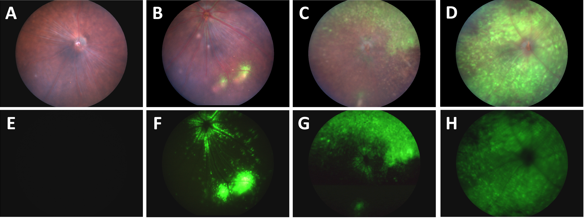
In their article, “A Novel Method Combining Vitreous Aspiration and Intravitreal AAV2/8 Injection Results in Retina-Wide Transduction in Adult Mice,” Da Costa et al use the Phoenix MICRON® III imaging platform to take stunning images demonstrating the success of their novel intravitreal injection technique. Gene therapy is a promising treatment option of genetic retinopathies—adeno-associated viruses […]
18.11
2020
The Phoenix MICRON® IV system and OCT help evaluate promising compounds for treatment of age-related macular degeneration
Age-related macular degeneration affects tens of millions of people worldwide, leading to vision impairment and blindness. Anti-VEGF treatment helps only 25-40% of patients, leaving others with no recourse to this progressive blinding disease. In their article, “Suppression of aberrant choroidal neovascularization through activation of the aryl hydrocarbon receptor,” Choudhary et al explore potential treatment using […]
21.07
2020
Caspase-9 inhibiting eyedrops rescue physiological and functional retinal vein occlusion damage shown with Phoenix MICRON®, OCT, and focal ERG
In a recent well written, compelling article published in Nature Communications, “Endothelial activation of caspase-9 promotes neurovascular injury in retinal vein occlusion,” Avrutsky et al show that caspase-9 inhibition is a promising treatment for retinal vein occlusion. Retinal vein occlusion models hypoxic-ischemic neurovascular damage and is the second leading cause of blindness in working-age adults. […]
23.06
2020
Targeting VEGF164 in Müller cells may be useful to treat retinopathy of prematurity
In Nature’s Scientific Reports, Becker et al use the Phoenix MICRON® IV, OCT, and focal ERG to assess the therapeutic value of knocking down a splice variant of VEGF in Müller cells in a model of Retinopathy of Prematurity (ROP). ROP is characterized by delayed vascularization of the retina after disrupted oxygen levels, followed by […]
20.05
2020
Imaging unanesthetized mice with the Phoenix MICRON® OCT provides consistent retinal degeneration measurements
In the recently published review, “Mouse Models of Inherited Retinal Degeneration with Photoreceptor Cell Loss,” researchers from The Jackson Laboratory in Bar Harbor, Maine and Mahidol University in Bangkok, Thailand performed an extensive literature search to find mouse models of single-gene mutations leading to photoreceptor loss and retinal degeneration. Using the Phoenix MICRON® III and […]


