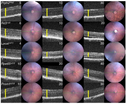
In the recently published review, “Mouse Models of Inherited Retinal Degeneration with Photoreceptor Cell Loss,” researchers from The Jackson Laboratory in Bar Harbor, Maine and Mahidol University in Bangkok, Thailand performed an extensive literature search to find mouse models of single-gene mutations leading to photoreceptor loss and retinal degeneration. Using the Phoenix MICRON® III and IV fundus camera and the Phoenix MICRON® OCT on unanesthetized mice, they documented the fundus appearance and retinal structure of selected mouse mutants. The crisp OCT images (Figure 1) reveal the varied uses of the Phoenix OCT—although sedated mice offer more opportunities for clear, averaged images, the OCT can capture informative images from non-sedated mice as well.
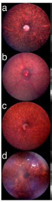
The review begins with an excellent summary of photoreceptor anatomy and function followed by a detailed description of the article search parameters in Mouse Genome Informatics and PubMed which found plentiful examples of single-gene mutations resulting in retinal degeneration with photoreceptor loss. The Eye Mutant Resource and the Translational Vision Research Models centers at The Jackson Laboratories image retired breeders to screen for ocular disease models. These mice were examined with the Phoenix MICRON® and Phoenix MICRON® OCT to enhance the information collected from the articles. The OCT images—collected on non-sedated mice, a tricky and impressive technical feat—show clear and varied degeneration patterns (Figure 1). The Phoenix MICRON® fundus images, taken from sedated animals, also reveal abnormalities (Figure 2).
Synthesizing the image and article data, Collin et al provide a detailed analysis of the genetic data with possible gene function and gene interactions. They analyzed the genes to create functional clusters and noted photoreceptor loss patterns among these clusters, then earmarked potential models of human disease. This analysis generated an extensive list of mouse models of single-gene retinal degeneration disease and an updated Mouse Genome Informatics, a database with expert annotation from The Jackson Laboratories, to encourage further research. Inherited retinal degeneration diseases such as retinitis pigmentosa, Leber congenital amaurosis, Usher syndrome, etc result in devastating impaired vision or blindness due to photoreceptor loss. This article may contribute to treatments and cures by providing a guide to consistent measurement and a number of new mouse models for human disease.
Figure 2. Phoenix MICRON® images of retinal degeneration through photoreceptor loss mouse models from the Translational Vision Research Models at The Jackson Laboratory. Images taken on sedated mice with dilated eyes, providing a slightly clearer look at the fundus
Citation:
Collin, G. B., Gogna, N., Chang, B., Damkham, N., Pinkney, J., Hyde, L. F., Stone, L., Naggert, J. K., Nishina, P. M., & Krebs, M. P. (2020). Mouse Models of Inherited Retinal Degeneration with Photoreceptor Cell Loss. Cells, 9(4), 931. https://doi.org/10.3390/cells9040931


