In their 2017 article, “Effect of subretinal injection on retinal structure and function in a rat oxygen-induced retinopathy model,” Becker et al used the Phoenix MICRON® IV fundus camera, Phoenix MICRON® OCT2 and corresponding layer analysis software Insight 2D, and the Phoenix MICRON® focal ERG to find that subretinal injection of saline or even introduction […]
28.10
2020
A year-long longitudinal pattern dystrophy fundus study with the Phoenix MICRON® IV imaging platform
In their 2019 paper, “Novel molecular mechanisms for Prph2‐associated pattern dystrophy,” Chakraborty et al use the Phoenix MICRON® IV retinal imaging platform to longitudinally study the effect of a very specific mutation affecting the Peripherin 2 protein. Peripherin 2 is a protein in rods and cones which, if mutated, can lead to retinitis pigmentosa, cone-rod […]
26.08
2020
Phoenix MICRON® III shows microglia-like cells migrating from the optic nerve after injury
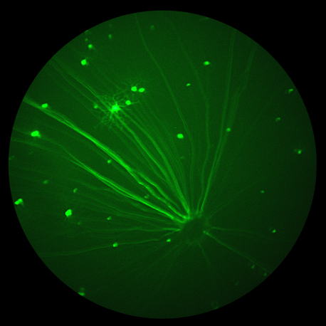
Microglia respond to neurological injury but the precise way they help to clear and remodel the injuries is not known. In their paper, “Optic nerve as a source of activated retinal microglia post-injury,” Heuss et al investigate a population of microglia-like cells that proliferate in the retina after an optic nerve injury. They identify GFPhi myeloid […]
21.07
2020
Caspase-9 inhibiting eyedrops rescue physiological and functional retinal vein occlusion damage shown with Phoenix MICRON®, OCT, and focal ERG
In a recent well written, compelling article published in Nature Communications, “Endothelial activation of caspase-9 promotes neurovascular injury in retinal vein occlusion,” Avrutsky et al show that caspase-9 inhibition is a promising treatment for retinal vein occlusion. Retinal vein occlusion models hypoxic-ischemic neurovascular damage and is the second leading cause of blindness in working-age adults. […]
20.09
2019
Phoenix MICRON® OCT tracks individual stem cells in the rat retina
In the July edition of Nanomedicine journal, Chemla et al demonstrate a fascinating and novel way to label and track individual photoreceptor precursor cells migrating within the retina with fluorescence and gold nanoparticle tagging using the Phoenix MICRON® and OCT. Many retinal diseases such as age-related macular degeneration and retinitis pigmentosa are characterized by photoreceptor […]
16.01
2018
Using the MICRON® IV to Study Light Induced Retinal Degeneration
Dr. Rafael Ufret-Vincenty’s lab at University of Texas Southwestern Medical Center has developed a novel model for light damage using the Micron IV rodent retinal imaging camera. This quick and consistent light damage model leads to fundus abnormalities and retinal thinning as measured by the Micron image-guided OCT and semi-automated layer analysis tool, Insight. In two elegant articles, the researchers provided proof of concept in pigmented mice, which are a better model for human eye light damage than overly sensitive albino mice, which demonstrated bleached fundus and outer retinal layer thinning.


