Microglia respond to neurological injury but the precise way they help to clear and remodel the injuries is not known. In their paper, “Optic nerve as a source of activated retinal microglia post-injury,” Heuss et al investigate a population of microglia-like cells that proliferate in the retina after an optic nerve injury. They identify GFPhi myeloid cells in the CD11cGFP mouse that dynamically respond to injury, specifically contacting the injured areas and acting as dendritic cells in a manner distinct from GFPlo resident retinal microglia. Their paper characterizes these cells through many clever and complex experiments involving layers of fluorescent cells, parabiotic mice, Tam-induced RFP-expression, and much flow cytometry and fluorescent RFP and GFP fundus imaging with the Phoenix MICRON® III.
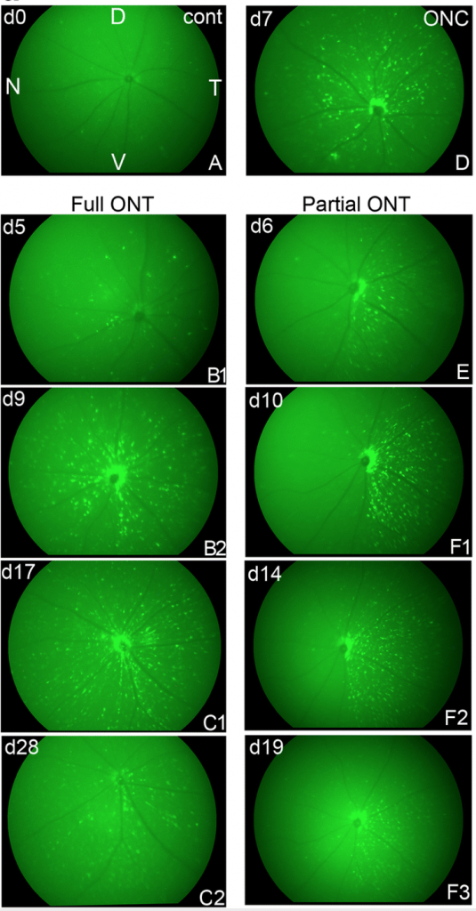
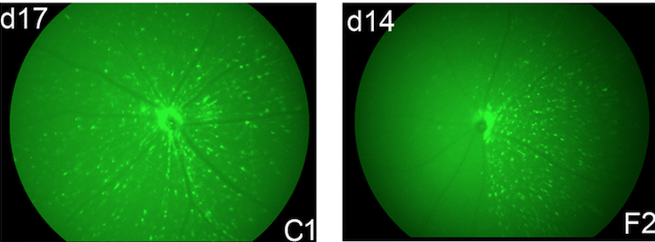
Heuss et al determined that the GFPhi myeloid cells were not recruited from circulation but instead generated from optic nerve microglia and migrate along the optic nerve fibers into the retina after an injury. The Phoenix MICRON® III revealed stunning images of these GFPhi myeloid cells responding to optic nerve crush or transection in a distinct pattern in the retina. The researchers used partial and full optic nerve transections to recruit the GFPhi myeloid cells to the retina. In an uninjured mouse (Figure 1 A), there are no GFPhi myeloid cells. In an optic nerve crush (Figure 1 D), the GFPhi myeloid cells are randomly dispersed. With a full optic nerve transection, the GFPhi myeloid cells radiate from the optic nerve and migrate into the retina over the course of several days (Figure 1 B1-C2), whereas with a partial nerve transection (Figure 1 E-F3), the GFPhi myeloid cells only migrate into a select wedge of the retina, presumably crawling along the damaged axons.
The article uses a range of innovative techniques to characterize a distinct population of injury-activated cells. The live retinal fluorescent imaging as well as the various fluorescent histology is impressive and collected into a stunning collection of images.
Heuss, N. D., Pierson, M. J., Roehrich, H., McPherson, S. W., Gram, A. L., Li, L., & Gregerson, D. S. (2018). Optic nerve as a source of activated retinal microglia post-injury. Acta Neuropathologica Communications, 6(1), 66. https://doi.org/10.1186/s40478-018-0571-8