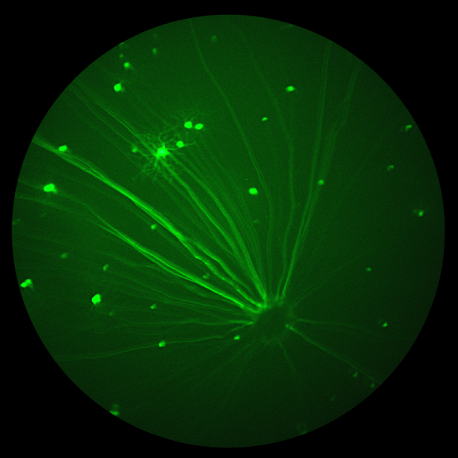
In the paper “The impact of obesity‑related raised intracranial pressure in rodents” published in Scientific Reports journal, Westgate and collaborators used the Phoenix MICRON® image-guided OCT system, along with the Phoenix MICRON® Insight software, in the measurement of the retinal anatomical changes caused by intracranial hypertension in obese rats. The Problem: Intracranial pressure (ICP) measures the […]
28.02
2022
Analysis of imaging, structure and function using Phoenix MICRON™ modalities expand the understanding of ocular features of Down Syndrome in mouse models
In their paper “Quantitative Analysis of Retinal Structure and Function in Two Chromosomally Altered Mouse Models of Down Syndrome”, researchers Victorino, Scott-McKean, et al leveraged the multi-modality capabilities of the Phoenix MICRON™ retinal imaging platform, to produce an image-rich research paper looking at the ocular features of Down Syndrome in two mouse models; Ts65Dn and […]
19.10
2021
Cataracts caused by lens protein knockout imaged using the Phoenix MICRON® IV Slit Lamp
In their article, “CRYβA3/A1-Crystallin Knockout Develops Nuclear Cataract and Causes Impaired Lysosomal Cargo Clearance and Calpain Activation,” Hegde et al use the Phoenix MICRON® IV Slit Lamp to examine the effect of knocking out a lens structural protein. α, β and γ crystallins are lens structural proteins that are needed for transparency and refractive power […]


