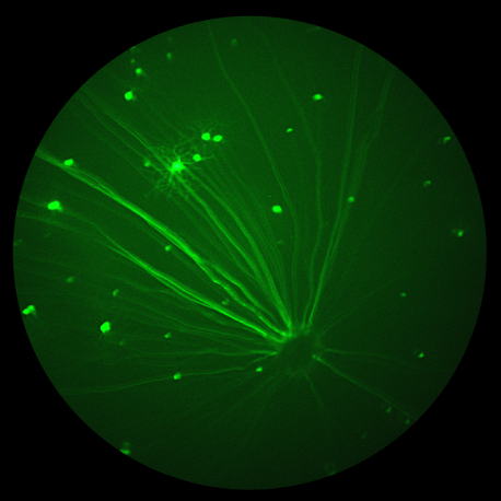
Phoenix extends its product offerings in North America to include behavioral assays to evaluate the visual abilities of rodents Bend, OR, USA, January 25, 2024 – Phoenix-Micron, Inc. (“Phoenix”), recognized globally for its leadership in in vivo ophthalmic imaging of small animals, is proud to unveil its latest collaboration as the exclusive North American […]
Phoenix MICRON Research Blog
12.12
2023
Exploring the Intersection of Art and Retinal Research: A Journey of Discovery
Our latest blog post veers from the traditional focus on a singular research paper to bring you an enthralling narrative at the crossroads of progressive inherited retinal dystrophy (IRD), the frigid expanses of Antarctica, marathon endurance, the expressive world of art, and the transformative realm of retinal research. This story encompasses gene editing, animal models, […]
16.08
2023
Small zebrafish are impressive, effective models for studying endotoxin induced uveitis
Researchers at Henan Province Eye Hospital in China have made great strides in studying endotoxin-induced uveitis (EIU) by creating a novel EIU model in zebrafish. Uveitis is an ocular inflammation and one of the main causes of visual impairment, accounting for 10-15% of global blindness. Mice have typically been the research animal of choice however […]
11.07
2023
Researchers find early signs of Alzheimer’s disease (AD) in the mouse retina and develop a non-invasive artificial intelligence-based system that could be used to diagnose the disease before the appearance of clinical symptoms
Progress in diagnosing and treating Alzheimer’s disease (AD) has been accelerating over the last few years. Until now, AD has been difficult to diagnose. The disease can be present years before distinguishable symptoms manifest. Non-invasive diagnostic tests have been lacking, and diagnosis often relies on memory, cognitive, and behavioral tests. In 2011, Koronyo-Hamaoui M, Koronyo […]
21.04
2023
Phoenix-Micron, Inc. launches the next generation of its MICRON in vivo ophthalmic imaging platform for small animal research
The all-new MICRON® 5 platform delivers advanced performance, repeatability, and imaging workflow efficiency Bend, OR, USA, April 21, 2023 – Phoenix-Micron, Inc. today announced the launch of the all-new Phoenix MICRON® 5 imaging system, the latest breakthrough in small animal ophthalmic imaging technology. With over 450 published studies utilizing MICRON imaging systems, the MICRON platform […]
21.04
2023
Phoenix-Micron, Inc. Announces New, Comprehensive Imaging Software and Redesigned Lens Technology for the Phoenix MICRON® Imaging Platform
Two new innovations, MICRON Software Suite and MICRON LT2 lens technology, streamline the ophthalmic imaging workflow and deliver new data management capabilities. Bend, OR, USA, April 21, 2023– Phoenix-Micron, Inc., a leader in ophthalmic imaging technology, today announced the release of the new MICRON® Software Suite and the MICRON LT2 lens technology. The new […]


