Glaucoma, a leading cause of irreversible vision loss, is characterized by progressive damage to retinal ganglion cells (RGCs) and the optic nerve, often associated with increased intraocular pressure (IOP). While previous studies have implicated Tau protein expression and phosphorylation changes in other neurodegenerative diseases such as Alzheimer’s disease, Parkinson’s disease and glaucoma, the causative role […]
06.12
2017
Retinal Ganglion Cell Survival During Chronic and Acute Injury
Using the Phoenix retinal imaging and functional measurement to study retinal ganglion cell survival during chronic (glaucoma) and acute (optic nerve crush) injury
Liu et al studied the survival and dysfunction of retinal ganglion cells (RGC) during chronic (glaucoma) and acute (optic nerve crush) injury in a series of comprehensive and elegant articles published from 2015 to 2017. The researchers used the Phoenix ERG and Micron IV provide a complete picture of RGC disruption.
02.11
2017
Micron Reveals Decreased Retinal Ganglion Cell Arborization in a Mouse Model of Retinal Ischemia
Researchers Dailey et al, in the Mitton Lab at Oakland University used the Micron retinal imaging camera to examine retinal ganglion cell (RGC) survival in a mouse model of retinal ischemia. Oxygen-induced retinopathy (OIR) in mice recapitulates critical factors of the human diseases retinopathy of prematurity and diabetic retinopathy. Mice pups were raised in hyperoxegenated air (75% oxygen) for five days and then returned to room air (20% oxygen), which lead to pathological changes in the vascular and neural growth.
15.08
2017
Studying Glaucoma with Micron IV Fluorescent RGC Imaging
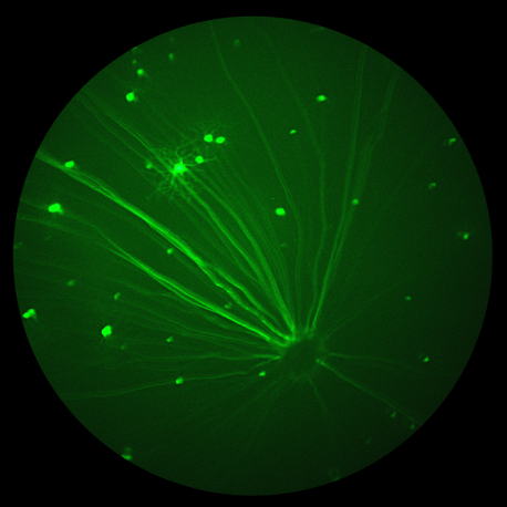
Dendrites may be retracted in several diseases as glaucoma. Studying morphology of dendritic arbors may give us an idea about functional deficits in those diseases. The Di Polo lab at the University of Montreal researches glaucoma using the Micron IV rodent retinal imaging camera and OCT module. Their scientists captured stunning fluorescent images with the Micron IV of mice genetically modified to produce yellow fluorescent protein (YFP)-taged retinal ganglion cells (RGC). the brightest RGC are visible in the bright field image along with blood vessels, optic nerve, and the retinal surface.


