Novel comparison of retina and brain vasculature leads to stunning Phoenix MICRON® images of fluorescent brain blood vessels
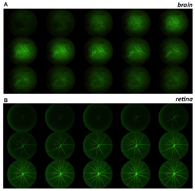
The Phoenix MICRON® imaging platform produces stunning images of fluorescent retinal vasculature through fluorescein angiography but for the first time, researchers have also captured beautiful images of cortical brain vasculature. Hui et al, in their well-written and engaging article, “Retinal and Cortical Blood Flow Dynamics Following Systemic Blood-Neural Barrier Disruption,” compared retinal and cortical response to fluorescein angiography and vasculature disruption. They shaved the skull, creating a window to the brain through which they could use the Phoenix MICRON® to image remarkable fluorescent vasculature after fluorescein injection (Figure 1A). These videos were compared with videos of retinal vasculature response to fluorescein (Figure 1B) to examine if retinal fluorescein angiography could be used to determine brain vasculature health. While there were some differences in fluorescein dynamics, the retina displayed blood-neural barrier disruption (leakage) before the brain did—confirming the clinical efficacy of using the retina as a representative for brain health.
Hui et al found some differences in fluorescein timing: potentially due to larger vessel size, the cortical vasculature filled with fluorescein and drained faster than retinal vessels and also had more residual fluorescein (Figure 2 shows analysis method). To compare retinal and brain response under disrupted conditions, the researchers administered sodium deoxycholate to disturb the brain blood barrier and retinal blood barrier. Interestingly, the retina showed leakage only 6 hours after administration while the brain took 24 hours to show leakage. Examining retinal fluorescein angiography is thus an excellent way to study the health of brain vasculature as it may show disruption even before the brain is affected. Imaging the retina vasculature can be a powerful tool to study brain health.
Hui F, Nguyen CTO, He Z, Vingrys AJ, Gurrell R, Fish RL and Bui BV. (2017). Retinal and Cortical Blood Flow Dynamics Following Systemic Blood-Neural Barrier Disruption. Front. Neurosci. 11:568.


