In their 2017 article, “Effect of subretinal injection on retinal structure and function in a rat oxygen-induced retinopathy model,” Becker et al used the Phoenix MICRON® IV fundus camera, Phoenix MICRON® OCT2 and corresponding layer analysis software Insight 2D, and the Phoenix MICRON® focal ERG to find that subretinal injection of saline or even introduction of a needle into the vitreal space has a detrimental effect on a rat model of retinopathy of prematurity. In normal adult rats, there is no effect of subretinal injections (results in other species vary) so these rats with oxygen-induced retinopathy may be more sensitive to damage.
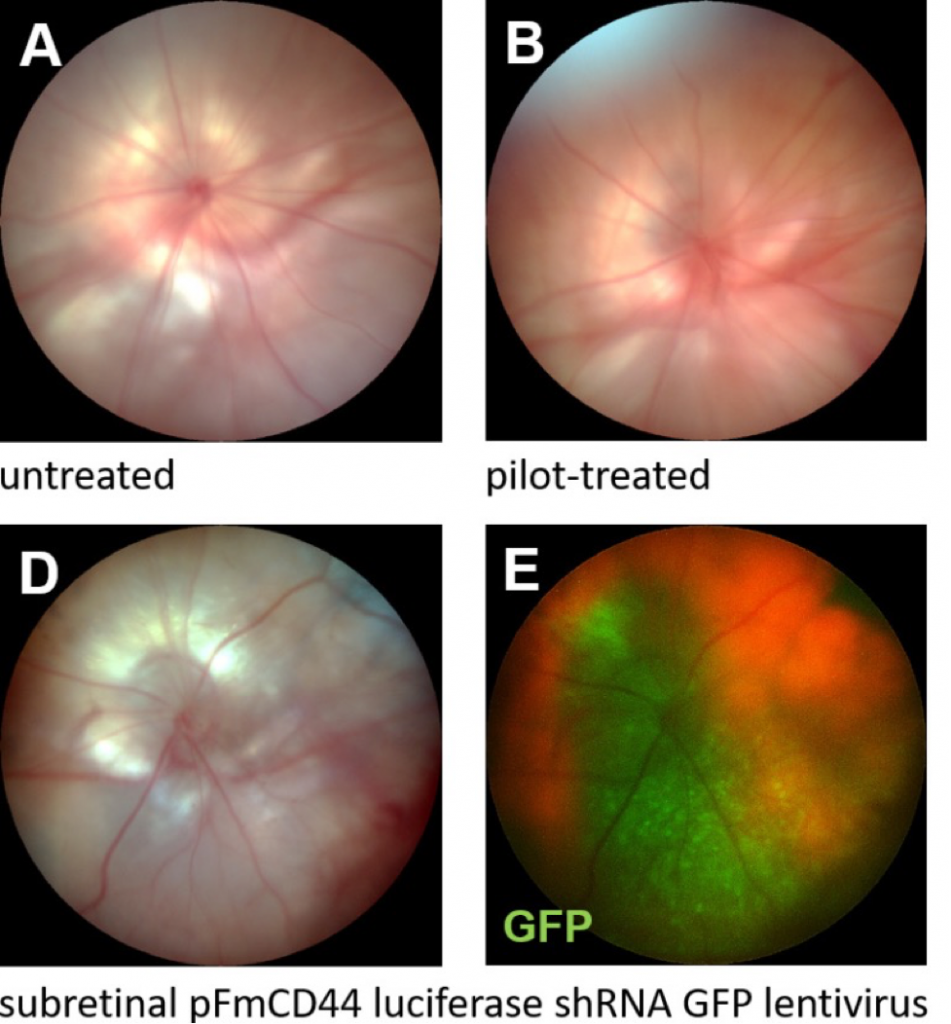
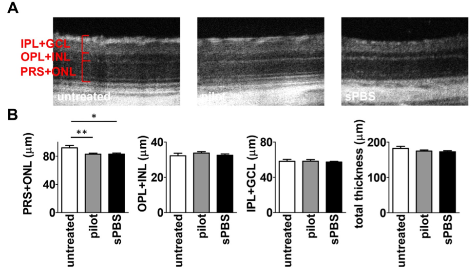
Becker et al induced a retinopathy of prematurity model in rat pups by oxygen cycling from birth through P14. At P8, they either subretinally injected PBS, introduced a needle into the vitreous without injecting anything (pilot-treated eyes), or did not inject eyes at all. They examined the fundus appearance with the Phoenix MICRON® IV and found no difference in appearance (Fig 1 shows the fundus appearance for oxygen-induced retinopathy rats as well as a GFP-expressing lentivirus for proof of concept). The Phoenix MICRON® IV OCT2 revealed reduced photoreceptor outer segment and outer nuclear layer thickness in both the PBS-injected and pilot-injected eyes (Fig 2A). The thickness was measured with the semi-automated Insight 2D OCT analysis software (Fig 2B).
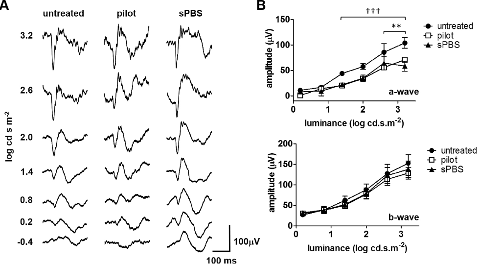
The Phoenix MICRON® focal ERG measured the electric response of the retina at the injection spot; since the focal ERG allows precise selection of stimulation area (under red illumination to maintain dark adaptation), Becker et al could identify and stimulate the injection site. The PBS-injected and pilot-injected eyes had reduced a-waves but normal b-waves (Fig 3). This aligns with the Phoenix MICRON® IV OCT findings that the photoreceptor layer was reduced.
Oxygen-induced retinopathy manifests as retinal thinning and reduced retinal function. Becker et al demonstrated that subretinal injection or even insertion of a needle into the vitreous cause more thinning and further reduced function. This has implications for considering treatment and when selecting control protocols in future studies.
Becker, S., Wang, H., Stoddard, G. J., & Hartnett, M. E. (2017). Effect of subretinal injection on retinal structure and function in a rat oxygen-induced retinopathy model. Molecular Vision, 23, 832–843.