Butterfly-shaped pigment dystrophy is an eye disease that produces butterfly-shaped lesions near the macula which can result in diminished visual acuity. In an article published in Nature Genetics, Saksens, Krebs, et al linked a mutation in the CTNNA1 gene to the disease in three families and found a mouse with the same mutation that showed a similar phenotype to the humans. The Micron IV rodent fundus camera revealed retinal dystrophy similar to humans.
In humans, severe butterfly-shaped pigment dystrophy manifests with an accumulation of pigmented material in the macula, diminished visual acuity, and abnormal eletro-oculogram recordings (Figure 2). It may progress to retinal atrophy, macular choroid disruption, and severe vision loss. The genetic basis was unknown until Saksens, Krebs, et al identified mutations in the CTNNA1 gene in three families with occurrences of the disease.
They then found mice with a similar genetic mutation (CTNNA1tvrm5 mice) and performed fundus imaging, OCT, and ERG to characterize the phenotype. As imaged by the Micron IV rodent fundus camera, the retina of homozygotes had a mottled appearance and bright spots, some with a dark center (Figure 1). The resolution of the Micron IV allows for close examination of the bright spots (Figure 1 c-e). Histology correlated with the bright spots shown in the fundus imaging, revealing pigmented cells accumulating on the apical surface of the RFE.
The OCT showed localized thickening and distortion of the photoreceptor inner-outer segment junction and external limiting membrane (Figure 3). Ring spots had an elevated, hyporeflective core surrounded by localized thickening. The ERG revealed significantly reduced scotopic a-wave and b-wave amplitudes in the homozygotes. More details can be found in the article.
Article citation: Saksens NT, Krebs MP, Schoenmaker-Koller FE, Hicks W, Yu M, Shi L, Rowe L, Collin GB, Charette JR, Letteboer SJ, Neveling K, van Moorsel TW, Abu-Ltaif S, De Baere E, Walraedt S, Banfi S, Simonelli F, Cremers FP, Boon CJ, Roepman R, Leroy BP, Peachey NS, Hoyng CB, Nishina PM, den Hollander AI. Mutations in CTNNA1 cause butterfly-shaped pigment dystrophy and perturbed retinal pigment epithelium integrity. Nat Genet. 2016 Feb;48(2):144-51. doi: 10.1038/ng.3474. Epub 2015 Dec 21.
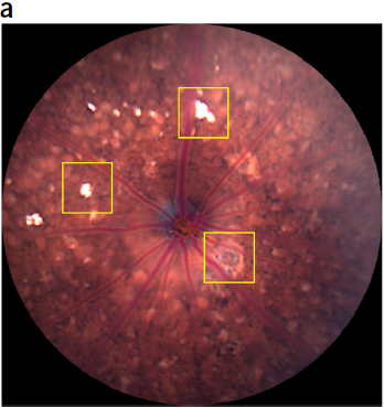
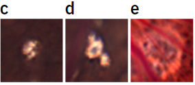
Figure 1. Ctnna1tvrm5 mice exhibit fundus mottling (a), spot lesions (c, d), and ring spot lesions (e). Images taken from live, sedated mice with the Phoenix Tech Group Micron IV, producing detailed, crisp fundus pictures.
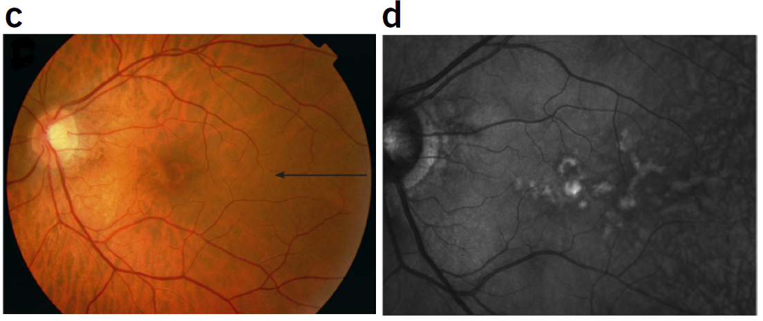
Figure 2. Examples of the human butterfly-shaped pigment dystrophy phenotype with mottled fundus (c) and hyper-reflective spots (d).
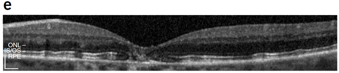
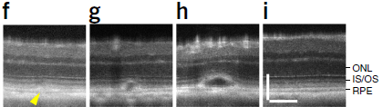
Figure 3. The human butterfly-shaped pigment dystrophy OCT (e) and mouse oct (f-i) show similar disruption.


