Hampel et al use the Phoenix MICRON® III imaging platform in an unusual manner in their paper “Relaxin 2 is functional at the ocular surface and promotes corneal wound healing by visualizing enucleated corneas.” The Phoenix MICRON® is designed to take crisp images and videos of the rodent retina, but this article demonstrates the other uses and creative methods of researchers using the MICRON®!
The article seeks to answer the question of if there is relaxin 2 present in the anterior segment of the eye and if relaxin 2 promotes wound healing. As ocular surface injuries are the most common problems seen by ophthalmologists, effective ocular surface wound healing that maintains the structure and clarity of the cornea is extremely important. Though relaxin is mostly known as a pregnancy hormone, it is present in various tissues and promotes extracellular matrix remodeling and thus affects wound healing. Hampel et al found that relaxin 2 was present in all ocular surface tissues analyzed and in human tears.
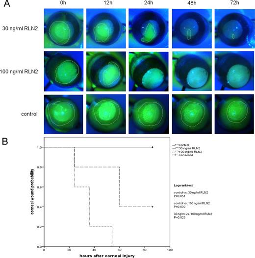
Hampel et al induced wounds in female C57Bl/6 mice by applying an alkaline mixture to their corneas for 2 minutes. They allowed the wounds to heal for 6 hours and then enucleated the corneas. The corneas were treated with control, a low relaxin 2 dose, or a high relaxin 2 dose and monitored every 6 hours for 84 hours. Wound healing was evaluated by using fluorescein eye drops and the included fluorescein imaging filters in the Phoenix MICRON® III camera (Fig 1 A). Fluorescein and GFP are easily visualized with a simple filter wheel turn switching from bright field to fluorescein filters in the Phoenix MICRON® camera.
Analysis was performed by circling the wound area and applying a Kaplan-Meier analysis (survival curve, Fig 1 B) with the endpoint of complete healing. As seen in Fig 1, the untreated corneas showed improvement but did not heal completely after 84 hours. 40% of the low dose corneas healed completely by 84 hours and 100% of the high dose corneas healed after 54 hours.
In conclusion, this study shows a mechanism for ocular surface wound healing and a promising treatment of applying relaxin 2. The methodology is compelling by revealing a novel and creative use of the Phoenix MICRON® III eye imaging camera.
Citation: Hampel, U., Klonisch, T., Makrantonaki, E., Sel, S., Schulze, U., Garreis, F., Seltmann, H., Zouboulis, C. C., & Paulsen, F. P. (2012). Relaxin 2 is functional at the ocular surface and promotes corneal wound healing. Investigative Ophthalmology & Visual Science, 53(12), 7780–7790.