Bend, OR, USA, November 29, 2021 — The newly formed company, Phoenix-Micron, Inc., announced today it has completed the spin-out of the Phoenix MICRON® imaging platform from Phoenix Technology Group. This move is designed to increase focus and innovation in products designed to serve the eye and eye-brain research community.
The new company, Phoenix-Micron, Inc. will be led by former Phoenix Technology Group CEO, Scott Carr, who served that role since 2018. Bringing Phoenix MICRON knowledge, and experience with imaging products for researchers, the team at Phoenix-Micron includes core R&D, engineering, sales, product, and manufacturing team members from Phoenix Technology Group.
The leading in-vivo retinal imaging system for researchers, Phoenix MICRON was launched in 2007 and was the first product marketed and sold by Phoenix Research Labs, which later became Phoenix Technology Group. Since its initial launch, MICRON camera systems have been installed in hundreds of eye and eye-brain research labs in more than 25 countries.
“This is a big day for the Phoenix MICRON product line,” said Scott Carr, Phoenix-Micron, Inc. president. “By returning to our roots, the Phoenix-Micron team, supported by a passionate lead investor, are better positioned than ever to drive innovations to help researchers see and understand the eye in ways that accelerate the prevention and treatment of blindness-causing conditions and diseases.”
Phoenix MICRON in-vivo ophthalmic imaging systems can be found in 11 of the world’s top 12 research institutes. More than 320 published research papers include imaging and data collected using MICRON systems. The MICRON system is not only the premier in-vivo ophthalmic imaging system for rodents, it also brings together multiple imaging modalities into a compact footprint that is ideal for the research lab.
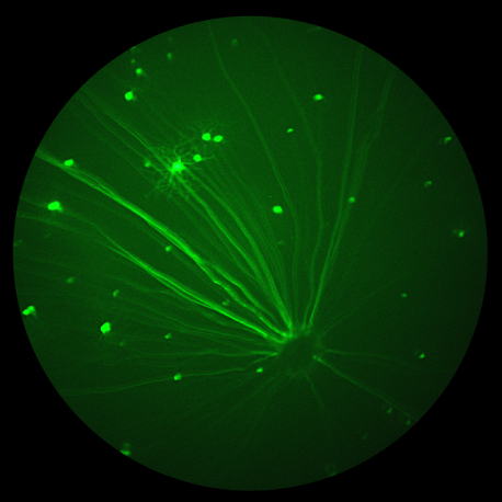
Today MICRON systems support bright field and fluorescent retinal and corneal imaging, stimulation of choroidal neovascularization, optical coherence tomography (OCT), and electroretinography (ERG). The combination of robust built-in features, analytic tools, and easy-to-add modalities coupled with stunning image quality make MICRON the system of choice for eye and eye-brain researchers.
“Phoenix MICRON started the in-vivo, retinal imaging category for rodents 14 years ago,” commented Al Klail, lead investor in Phoenix-Micron. “I’ve been a part of the company from almost the beginning, and I’m excited to be part of this next chapter. The MICRON name is synonymous with innovation and image quality. Our plan is to continue to sharpen our customer focus and invest to accelerate high-performance technology that supports researchers across ophthalmic and neuroscience research.”
About Phoenix-Micron, Inc.
Phoenix-Micron, Inc. provides the Phoenix MICRON® in-vivo ophthalmic imaging systems for eye and eye-brain researchers. Optimized for eye research using laboratory animals, both in computer simulations and in laboratory studies, Phoenix MICRON delivers performance at the physical limits of optical systems to fuel scientific discovery. Since its initial launch in 2007, MICRON camera systems have been installed in hundreds of ophthalmic and neurologic research labs in more than 25 countries and can be found in 11 of the world’s top 12 research institutes. MICRON systems provide ground-breaking eye and eye-brain research tools and have been used by authors of more than 320 published research papers. The MICRON product line includes support for bright field and fluorescent retinal and corneal imaging, stimulation of choroidal neovascularization, optical coherence tomography (OCT), and electroretinography (ERG). The combination of robust built-in features, analytic tools, and easy-to-add modalities coupled with stunning image quality and precise data capture make MICRON systems the platform of choice for leading researchers around the world.
For more information about Phoenix-Micron, Inc and the Phoenix MICRON® in-vivo imaging system, contact Jen King, jen.king@phoenixmicron.com.


