In Nature’s Scientific Reports, Becker et al use the Phoenix MICRON® IV, OCT, and focal ERG to assess the therapeutic value of knocking down a splice variant of VEGF in Müller cells in a model of Retinopathy of Prematurity (ROP). ROP is characterized by delayed vascularization of the retina after disrupted oxygen levels, followed by blood vessel growth into the intravitreal space, causing impaired vision and debilitating blindness Intravitreal anti-VEGF treats the vascularization but may also hurt delicate developing organs. Mouse studies using anti-VEGF treatment show smaller overall growth and reduced retinal capillary density as VEGF is important for both retina and organ development. A previous study targeting Müller cells with a short hairpin RNA against VEGF reduced intravitreal neovascularization without detrimental retina vascular effects, but also caused retinal thinning. The current study targeted the splice variant VEGF164 which is stimulated by oxygen fluctuations; mice without VEGF164 develop normally. The short hairpin lentivirus knocking down VEGF164 in Müller cells reduced intravitreal neovascularization and did not lead to retinal thinning.
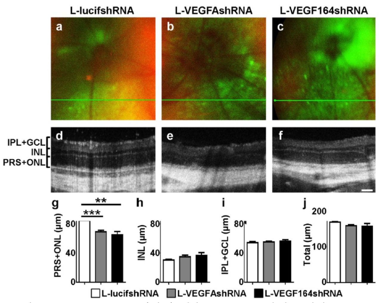
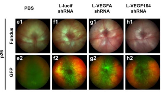
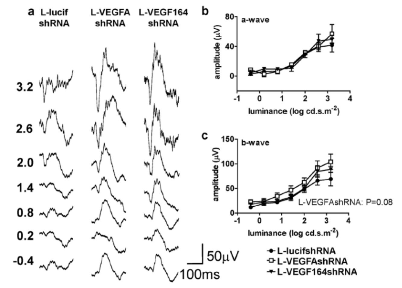
Rat pups were placed into a 10-50% oxygen cycling chamber for two weeks and then into room air to stimulate oxygen-induced retinopathy, a model of ROP. After one week in the oxygen-cycling chamber, one of three short hairpin lentiviruses with GFP targeting Müller cells was subretinally injected: one inert control, one VEGF knock down, and one VEGF164 knock down. The Phoenix MICRON® IV was used to quickly identify successful expression of the virus in-vivo via GFP expression (Fig 1). The Phoenix MICRON® OCT, which reveals the structure of the retina paired with live color video fundus, showed thinning of the photoreceptor and outer nuclear layers with knockdown of both VEGF and VEGF164, while the inner nuclear layer and the total retinal thickness unchanged. Insight 2D, a program included with the Phoenix MICRON® OCT, allows for easy semi-automated segmentation and retinal thickness measurements shown in Figure 2. The Phoenix MICRON® focal electroretinogram (ERG) allows for focal white light stimulus on a selected area of the retina. Using the focal ERG to measure the virus-expressing areas of the retina revealed no difference in function in the control, VEGF, or VEGF164 knockdown (Fig 3). While it is surprising that the VEGF knockdown did not harm retinal function, it may be because neuroprotective factors diffuse to the affected areas, and these were measured (Becker et al).
Using the Phoenix MICRON® system to supplement immunohistochemistry and mRNA analysis, Becker et al concluded that selectively knocking down VEGF164 in Müller cells may have fewer deleterious effects than nonselective VEGFA inhibition in all retinal cells
Becker, S., Wang, H., Simmons, A. B., Suwanmanee, T., Stoddard, G. J., Kafri, T., & Hartnett, M. E. (2018). Targeted Knockdown of Overexpressed VEGFA or VEGF164 in Müller cells maintains retinal function by triggering different signaling mechanisms. Scientific Reports, 8(1), 2003. https://doi.org/10.1038/s41598-018-20278-4


