Upgrade your research potential with the proven power of the MICRON® IV System
Versatile Scalability
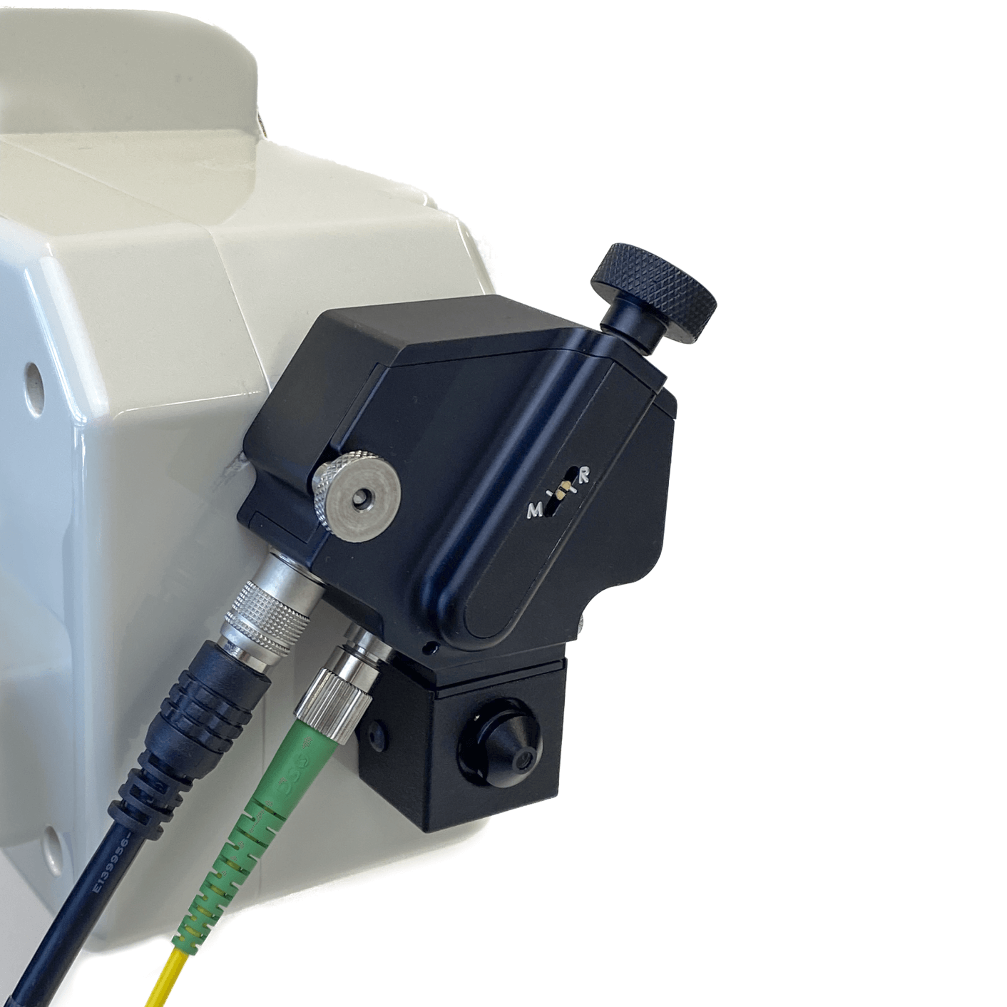
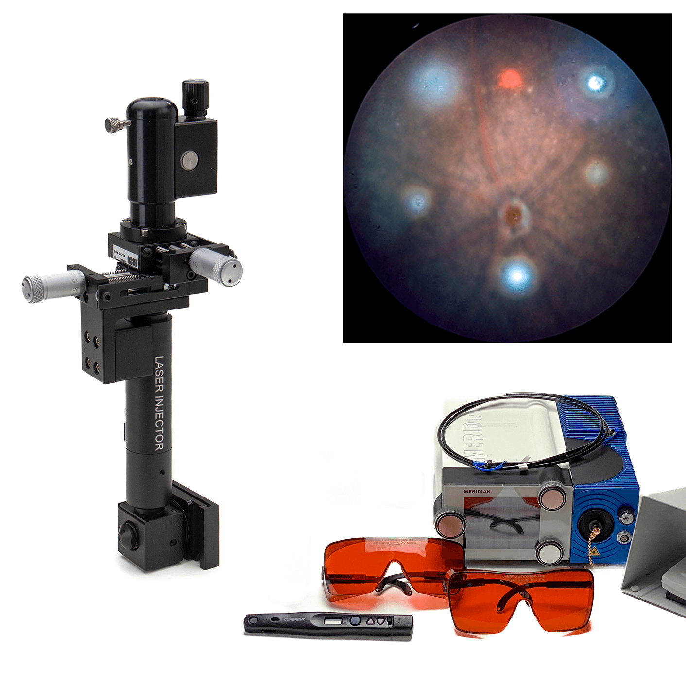
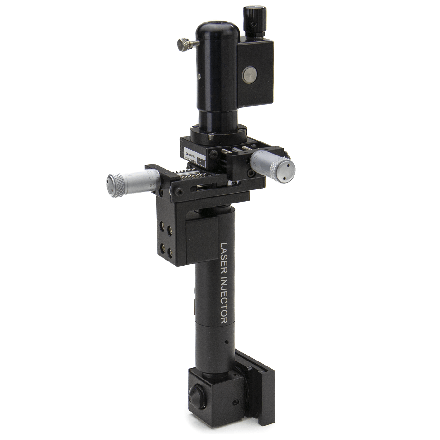
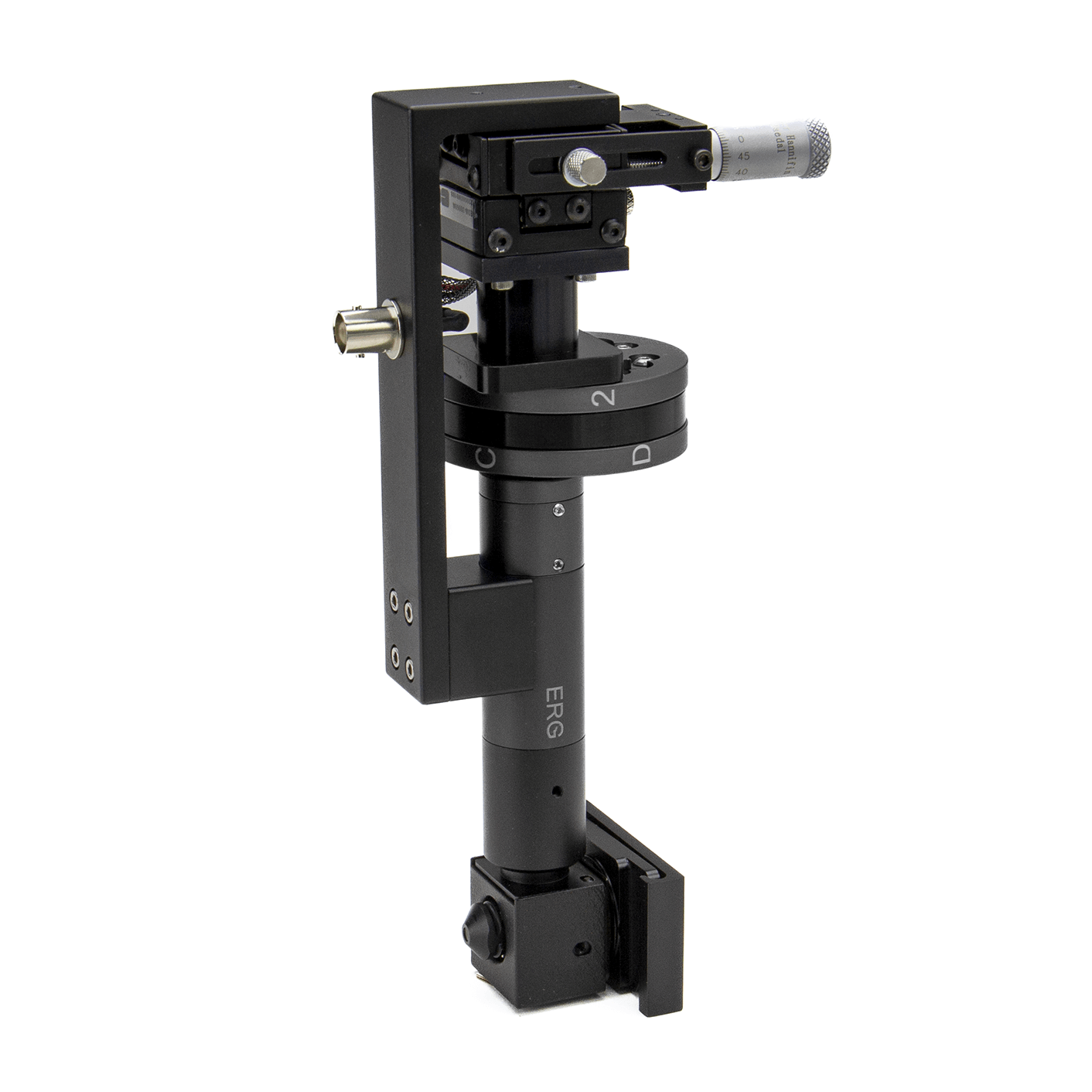
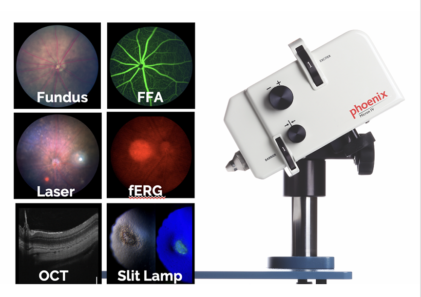
The MICRON® IV retinal microscope's scalable design allows researchers to expand its capabilities beyond the basic system. Although new MICRON® IV cameras are no longer available, researchers can still add Image-Guided OCT2, Image-Guided Laser, Image-Guided Focal ERG, or Slit Lamp modules to their existing MICRON® IV systems to increase research scope and leverage the efficiency of the small benchtop footprint.
Continued Support:
MICRON® IV systems continue to be widely supported and utilized in research centers across North America, Asia, and Europe.

Globally recognized
With over 900 research papers published using the system, the MICRON® IV is renowned for its exceptional core in vivo imaging capabilities for bright field, angiography, fluorescent, OCT, focal ERG and slit lamp imaging.

Extensible
Versatility makes the MICRON® an indispensable tool for a range of research applications, including basic eye research, toxicology, pharmaceutical efficacy, and neurological studies.

click on each tool below to learn more »
