MICRON® Geographic Atrophy System
Precise delivery of laser energy to the mouse and rat retina
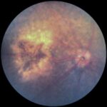
Preclinical research using rodent models is essential for advancing our understanding of dry age-related macular degeneration (AMD), a leading cause of vision loss with no approved cure. One powerful approach involves the use of an 810nm laser to induce geographic atrophy, enabling researchers to replicate key disease features and evaluate potential therapies in a controlled setting.
These models, combined with high-resolution imaging and analysis tools, are accelerating the development of treatments aimed at preserving vision and slowing disease progression.

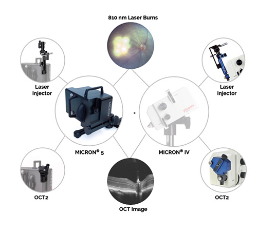
The MICRON® Geographic Atrophy System consists of:
- MICRON 5 imaging system
- Objective lens and laser injector attachment for 810nm laser
- 810 laser console and fiber optic
- Safety glasses
- Laser power meter
- MICRON Image-Guided OCT2 imaging modality
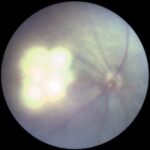
Real-time MICRON 5 imaging captures laser burns as they occur, while the combination of the 810 nm laser with fluorescein angiography (FA) and MICRON image-guided OCT with layer segmentation creates a powerful, end-to-end platform for preclinical in vivo research.


