Ground-breaking eye and eye-brain research tools used at hundreds of sites in 25 countries, and backed by over 320 research papers.
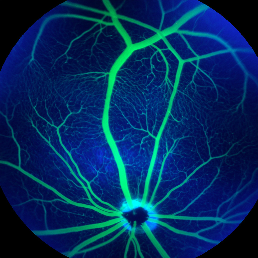
Optimized for eye research using laboratory animals, both in computer simulations and in laboratory studies, Phoenix MICRON® delivers performance at the physical limits of optical systems to fuel scientific discovery.
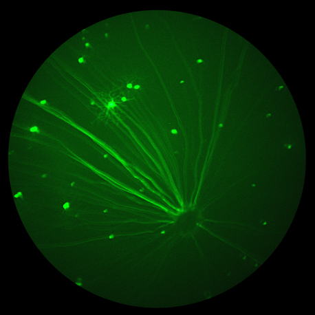
“Best rodent imaging system I’ve ever used. Produces exceptionally high quality digital fundus images of rodents and small animals. The resolution and contrast are much better than any other instrument we have tried. We have had good success with bright field, as well as fluorescence imaging of GFP expression in the retina and fluorescein angiography. The ability to capture video, select and output individual frames later is a big help when focusing on the retina in the small rodent eye.”
– John Flannery, PhD
Professor of Vision Science and Molecular and Cell Biology
Associate Director, Helen Wills Neuroscience Institute
University of California, Berkeley
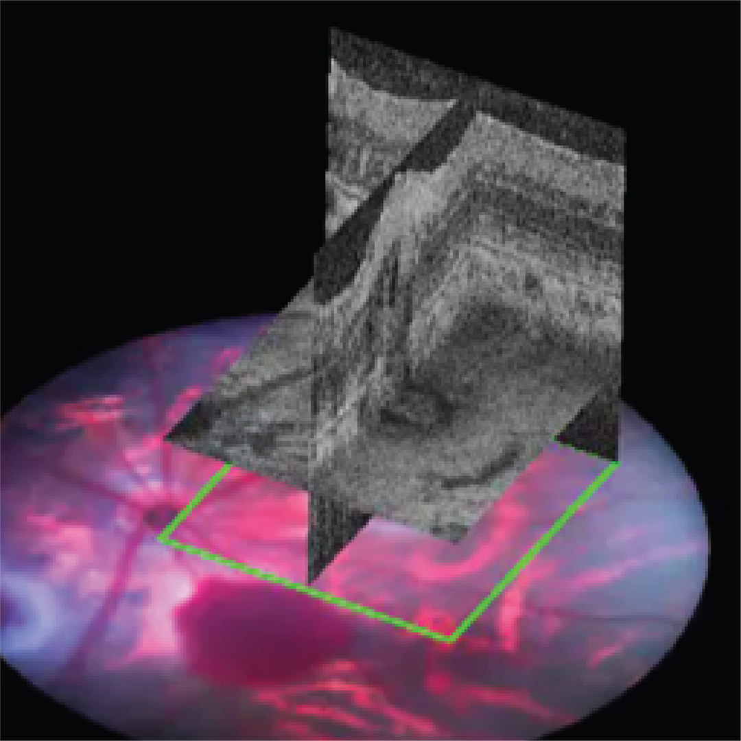
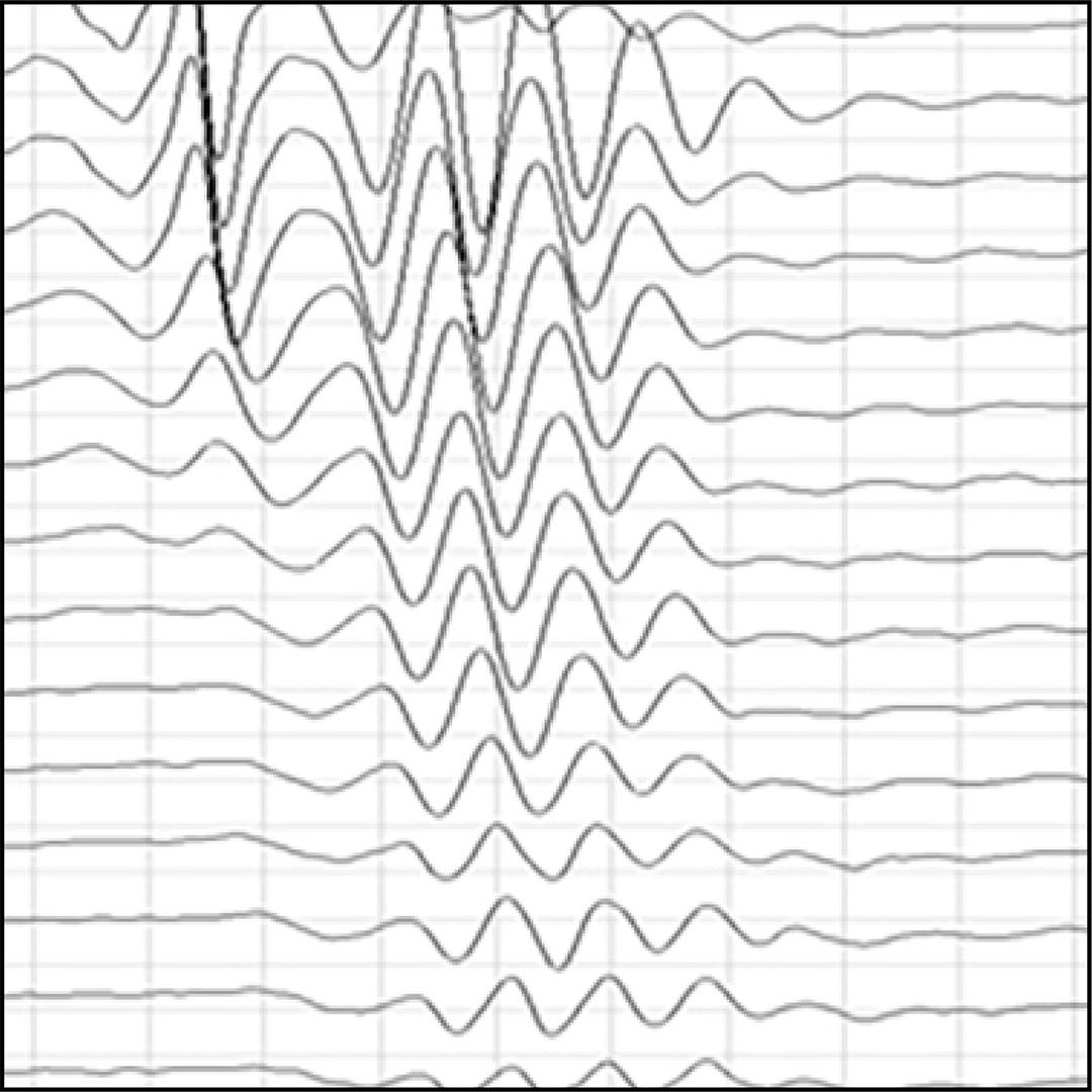
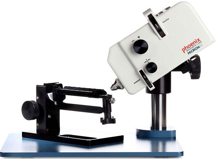
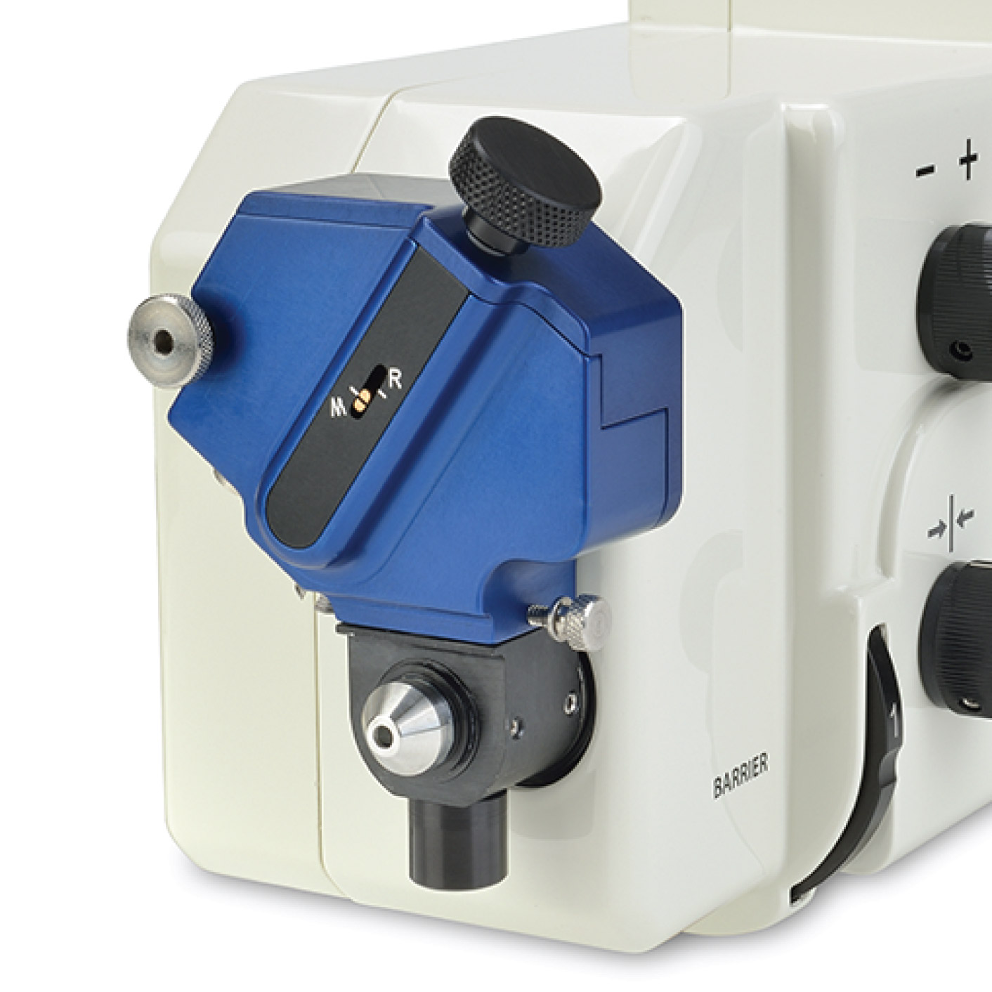
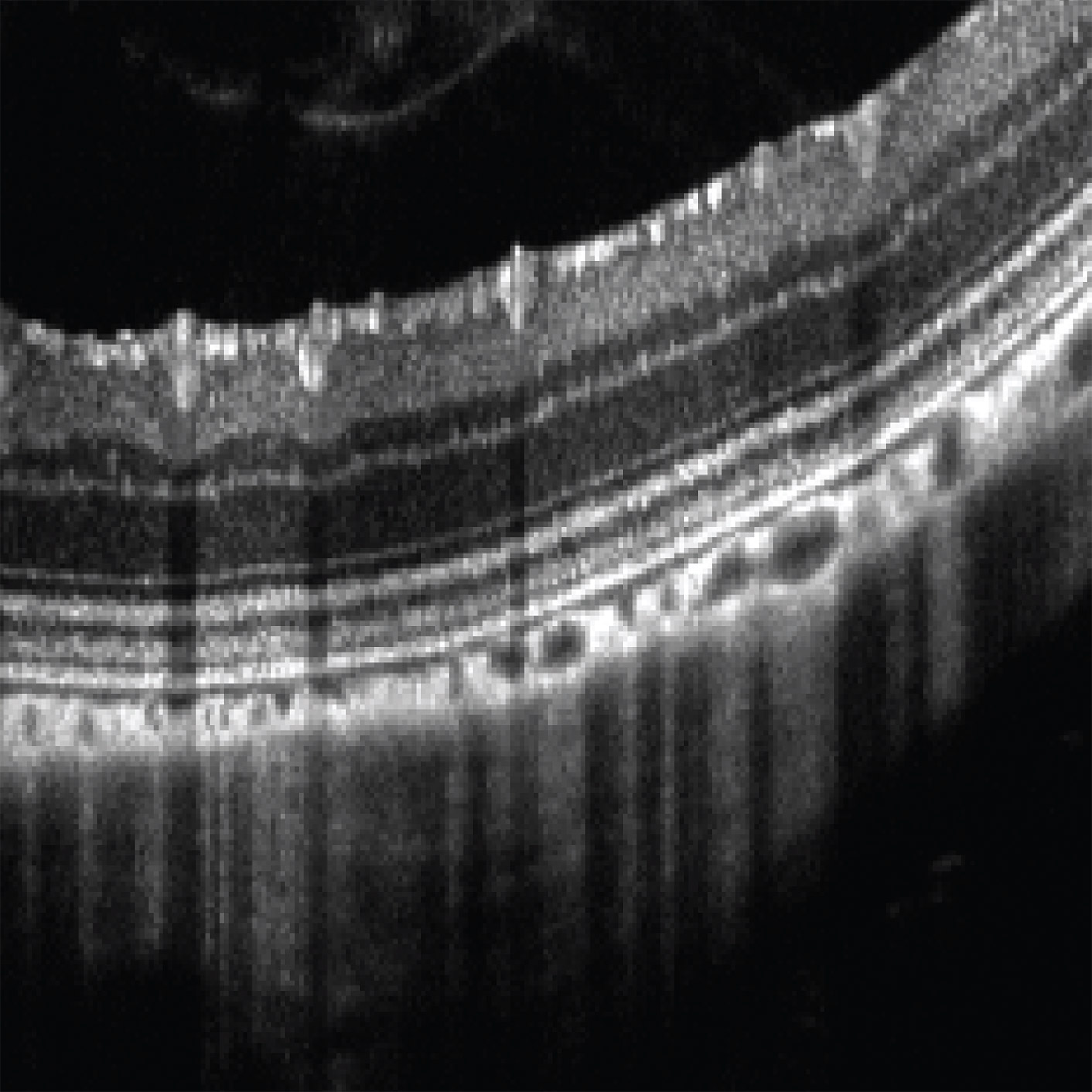
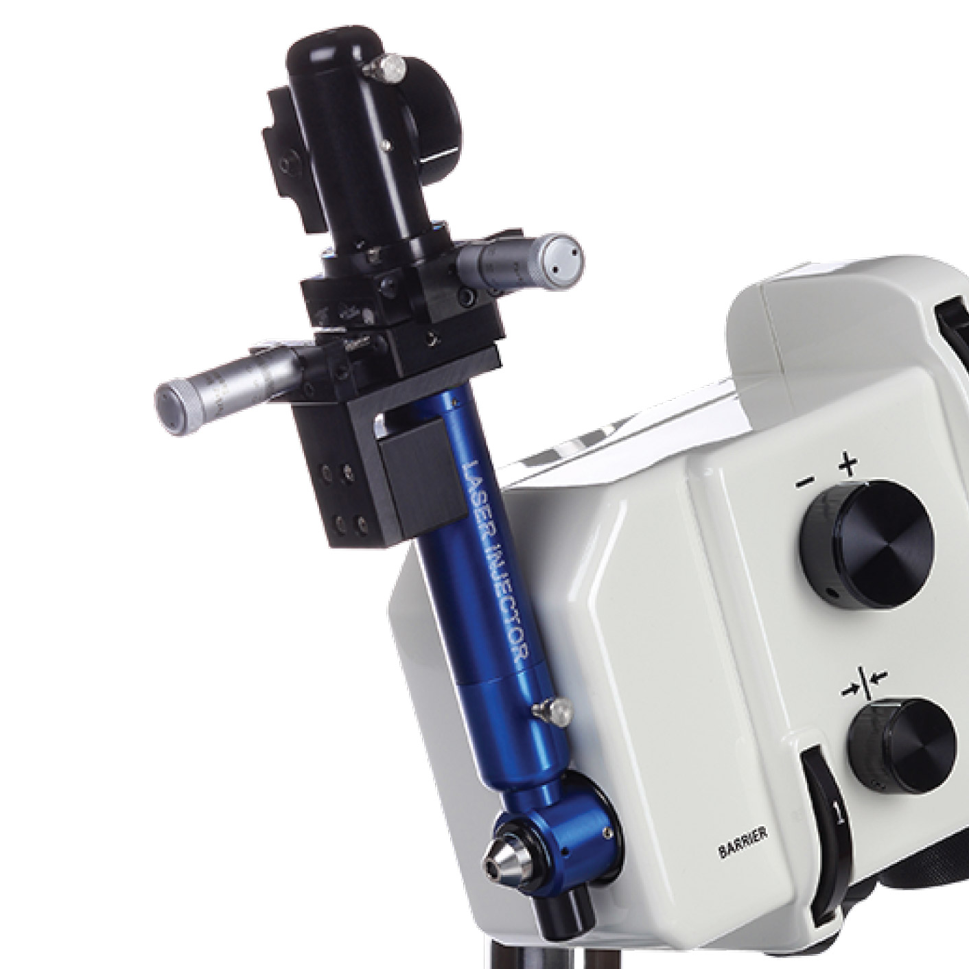
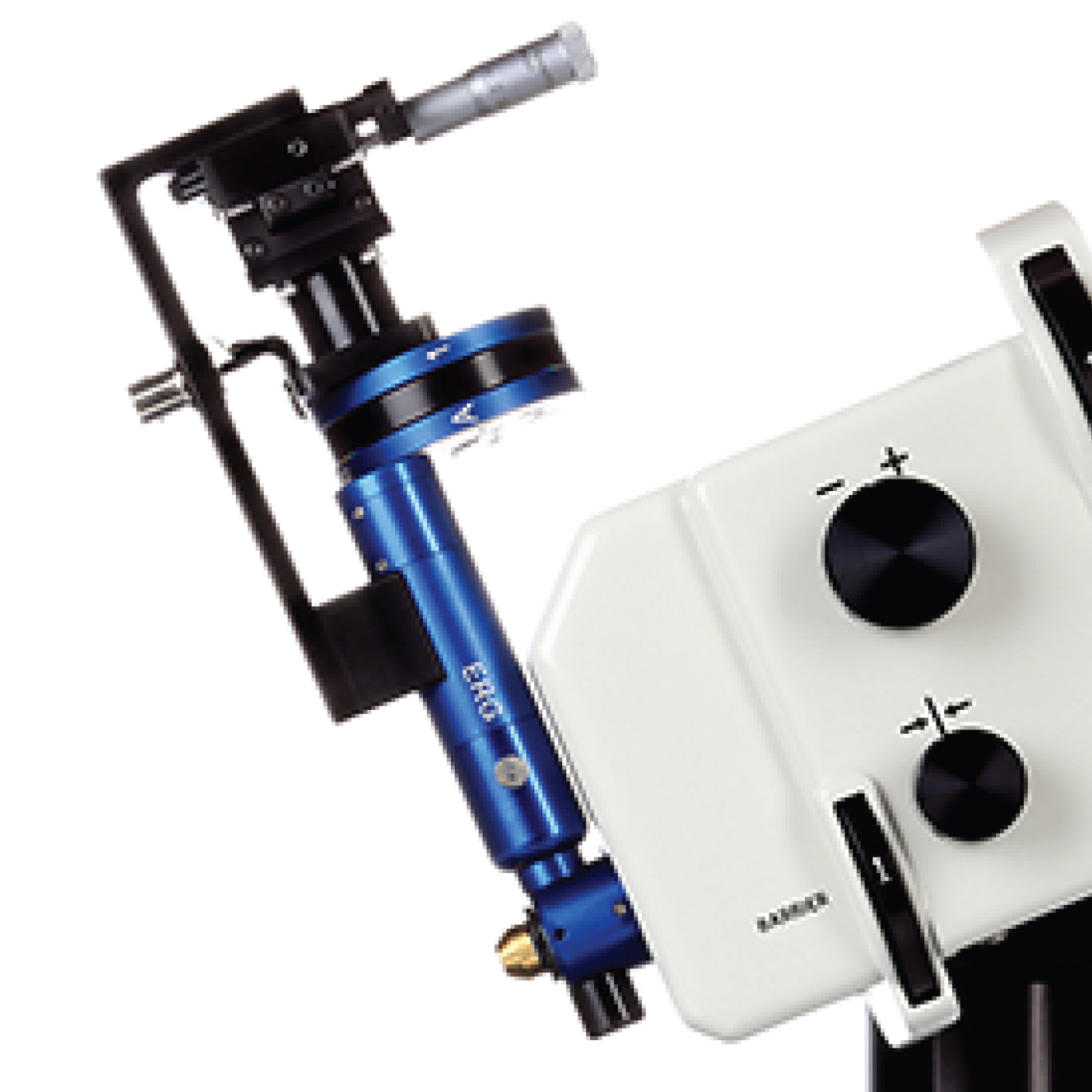
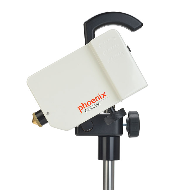
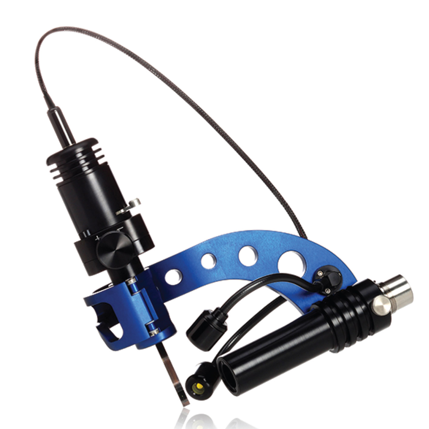
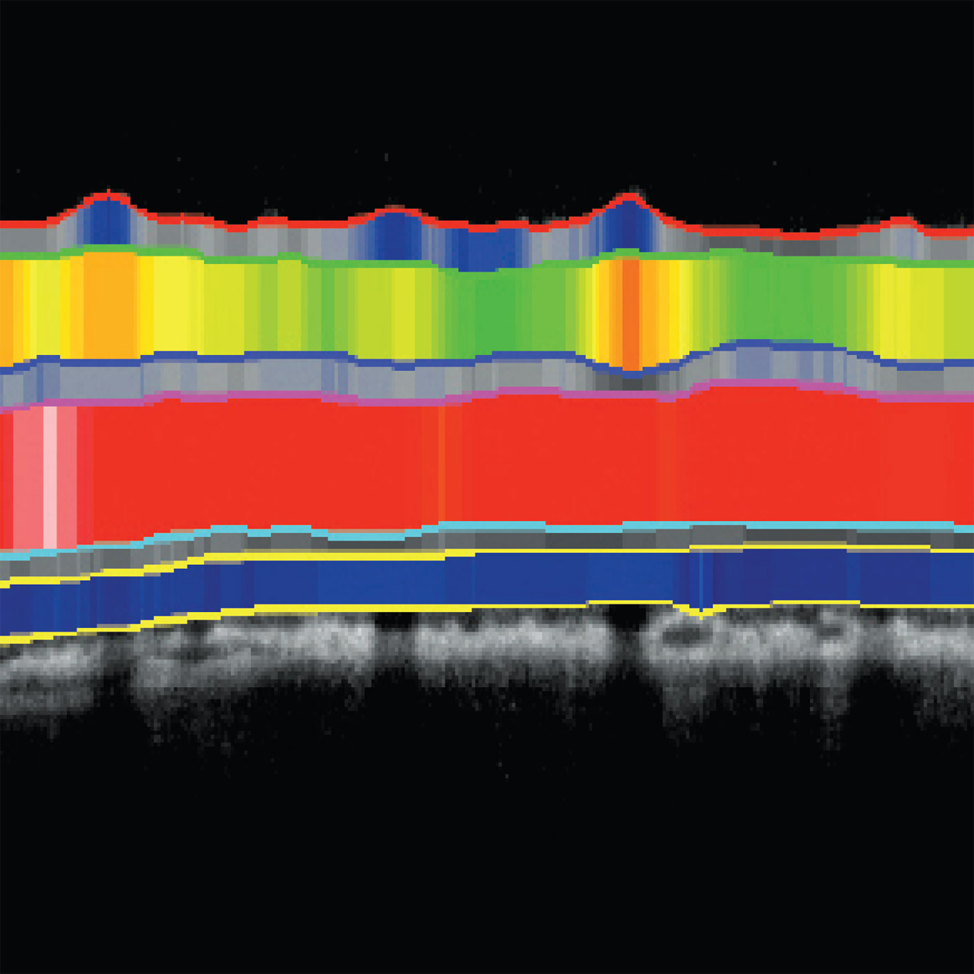
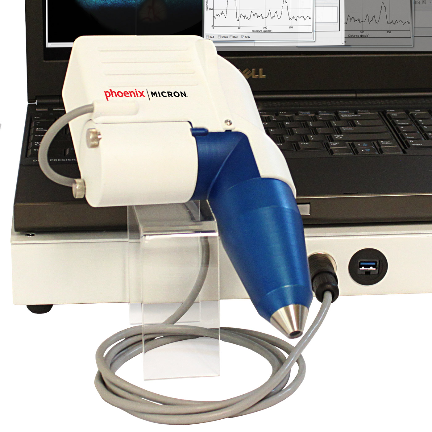
Our MICRON® platform features the Phoenix MICRON® IV Retinal Imaging Microscope for in vivo retinal imaging of small laboratory animals, and multiple add-on delivery tools to understand image, structure and function. Add-ons to the Phoenix MICRON® platform enable anterior segment imaging, laser injection, OCT and focal ERG.
The Phoenix MICRON® X2 is a unique portable retinal imaging system optimized for eye research using large animals.
click on each tool below to learn more »
