Another ARVO passed by in a blur of research, scientific discussions, and seeing science friends. If you came by our booth, thank you for swinging by to chat with the Phoenix team. If you didn’t get a chance, please let me know if you have any questions about the Micron system and look for our booth in Baltimore next year.
I enjoyed attending posters and talks given by a range of researchers. There was great research that used the Phoenix system. Here are some that stood out to me:
- Pushing the limits of retinal imaging: one clever researcher retrofitted the animal stand to fit parabiotic mice pairs to take retinal images with the Micron. Another took fundus images of a genetically modified frog!
- Advancing the study of age-related macular degeneration: The Phoenix laser remains a popular easy to use, consistent method to induce choroidal neovascularization to study the mechanisms—how an overactive immune response exacerbates the disease, for instance—and possible treatments for AMD
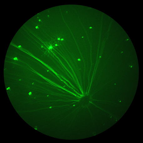
- Redefining “glue”: glia was named for glue since it was assumed to be unimportant support material for neurons—so many posters discussed the unexpected roles, positive and negative, that glia play in diseases and immune response. The images taken with the Micron of fluorescently-tagged glia that show the activated and unactivated states are beautiful.
- Immune responses in the eye: continuing a trend of the past several years, the immune system continues to play a role in seemingly everything. From showing that the periphery sends immune-activity to the eyes to showing that hampering the immune response can aid healing, research continues to flesh out a broad, intricate image of how the immune system hampers and helps achieve healthy eyes.
As always, please contact me if you have any questions about Phoenix, the Micron, or research. I’m happy to help with technique, experiment design, or even just see the results you’re getting with the Phoenix system!


