MICRON® Image-Guided OCT
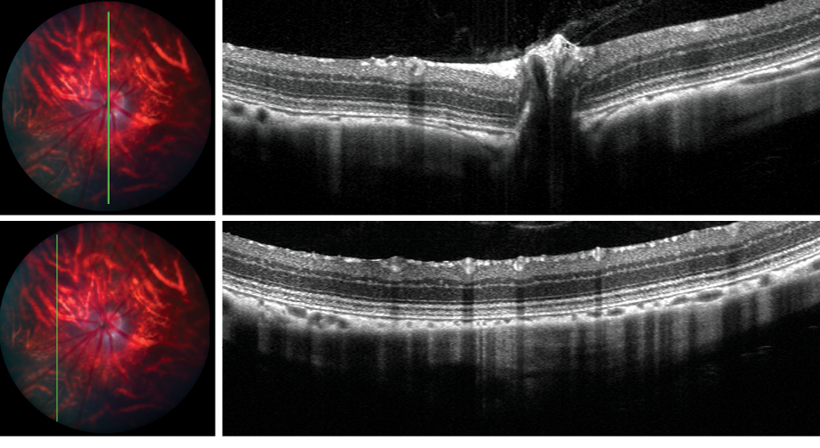
Stunning optical coherence tomography (OCT) optimized for mice and rats, with longitudinal resolution below 2 microns
MICRON Image-guided OCT:
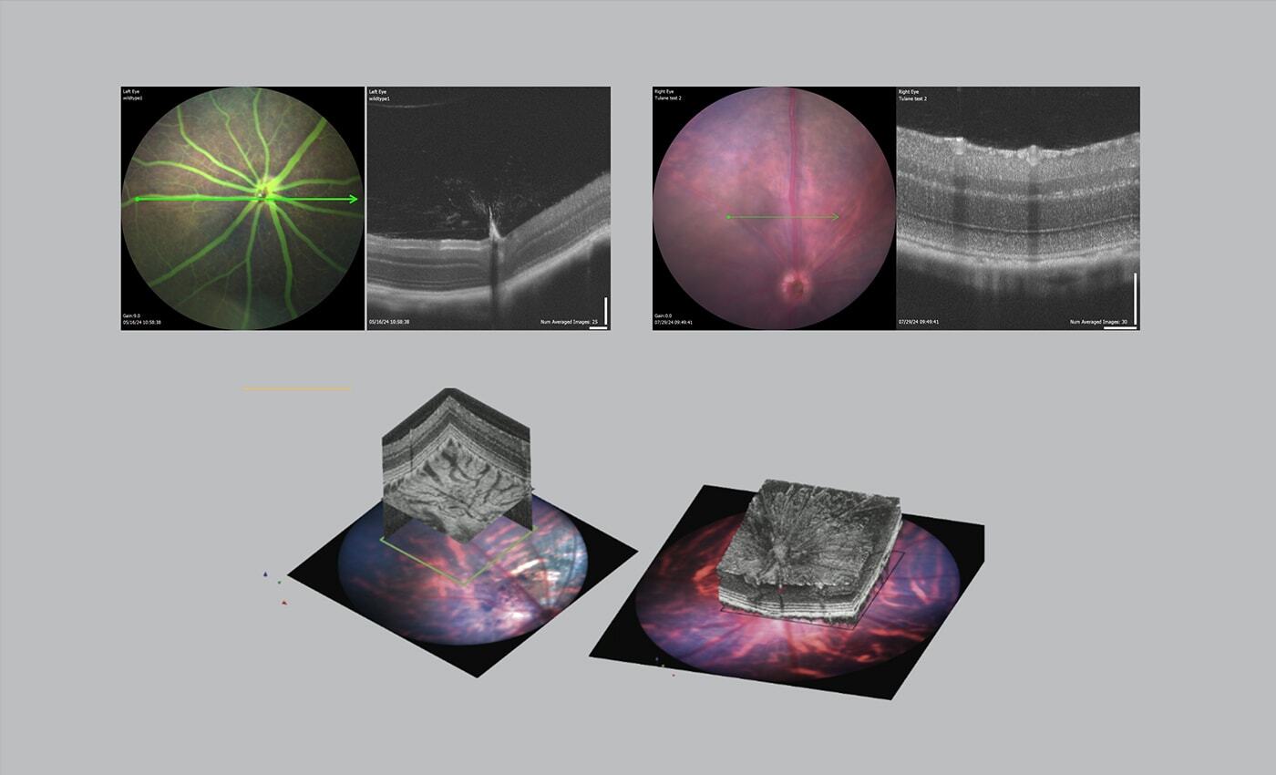
- Presents optical histological cross-sections on a real-time fundus view
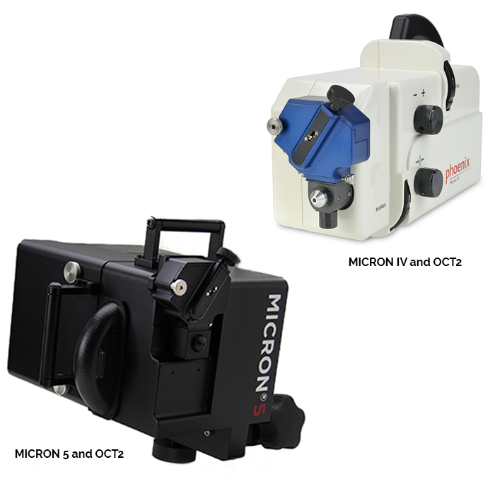
- Quickly attaches to the MICRON imaging system, and requires no additional bench space
- Reduces motion artifacts by imaging in contact with animal cornea
- Includes powerful 2D and 3D segmentation visualization tools to deliver retinal thickness values designed specifically for the mouse and rat eye

The MICRON® Image-Guided OCT2 system has been designed specifically for the demands of in vivo research using mice and rats, delivering stunning OCT images of mice and rats.
The OCT2 system includes a live, real-time fundus display with superimposed scan line to allow precise positioning of OCT imaging. The system supports 2D capture, and production of 3D volumes, and includes a set of powerful segmentation and visualization tools.
MICRON® Image-Guided OCT2 Features:
- Real-time, live fundus display alongside real-time OCT image presentation
- 1.8 micron longitudinal resolution
- 1.4 mm imaging depth, providing deep penetration into the sclera
- Line, circle, and 3D volume scan patterns
- Separate objective lenses optimized for mice and rats
- 2D and 3D segmentation tools, including interactive 3D visualization


