Image-Guided Laser
Photocoagulation
System

Color fundus image of a BALB/c mouse retina captured in vivo with the MICRON system, showing a translucent retina and visible choroidal vessels — features characteristic of albinism.

Color fundus image of a BALB/c mouse retina captured in vivo with the MICRON system, showing a translucent retina and visible choroidal vessels — features characteristic of albinism.

Fluorescein angiogram of a mouse retina captured in vivo with the MICRON system, highlighting large and fine vessels with uniform vascular filling and no signs of leakage or occlusion. The system's sensitivity enables visualization of blood flow, with individual cells often visible in motion during live video capture.

Retinal image of cyan fluorescent protein (CFP) expression captured with the MICRON system, showing fluorescent signal distributed through the retina and along the vasculature.

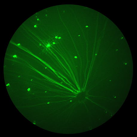
Fluorescent retinal image of GFP-labeled ganglion cells captured with the MICRON system, showing bright, discrete cell bodies scattered across the retinal field.

Color fundus image of a C57BL/6 wild-type mouse retina captured with the MICRON system, showing the optic nerve head with retinal vessels radiating over a healthy neurosensory retina.