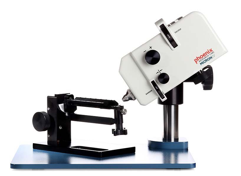
MICRON® MICRON® IV
Ophthalmic imaging systems for in vivo eye and eye-brain research using small animals.
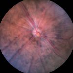
Designed to capture high-resolution, high-contrast retinal images and videos of mice, rats and other small animals. The MICRON IV delivers:
- Retina resolutions below 3 microns
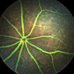
- Color fundus and fluorescent imaging, with interchangeable exciter and barrier filters
- Real-time display with capture of stills or videos
- Flexible & Scalable: Add Image-guided OCT, Image-guided Laser, Image-guided ERG, or Slit Lamp
“Exceptionally good imaging system for rodents. The whole system is user friendly, which makes it possible to finish the complete examination of an animal before any cataract can occur. The angiography function produces high quality images, where you can see single cells moving within the vessels. The use of this system to image mice with pigmented retinas is also absolutely possible..”
– Knut Stieger, DVM Ph.D.
Postdoctoral Research Fellow
Department of Ophthalmology
Justut-Liebig-University
Giessen, Germany
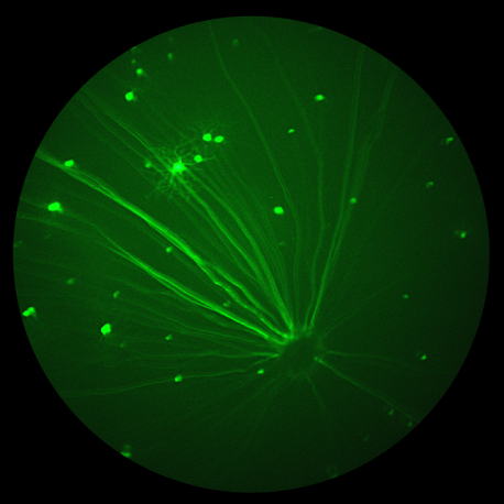
The Phoenix MICRON® IV retinal imaging microscope delivers remarkable bright-field images, fluorescein and Evan’s blue angiography, and fluorescent imaging of common reporter molecules such as GFP, YFP, mCherry and CFP.
By adding image-guided OCT, image-guided focal ERG, anterior segment imaging and image-guided laser delivery modules, you can expand the images available for your research quickly and easily.
Phoenix MICRON® IV Features:
- Custom three-chip CCD provides improved sensitivity for capturing even fainter fluorescent images.
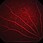
- New capability for imaging in the near infrared opens opportunities to capture long wavelength fluoropores and angiograms
- Improvements in the already easy-to-use ergonomic design enable additional filter choices and easier to use controls
