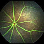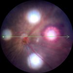
Image couleur du fond d'œil de la rétine d'une souris C57BL/6 de type sauvage capturée avec le système MICRON, montrant la tête du nerf optique avec des vaisseaux rétiniens rayonnant sur une rétine neurosensorielle saine.

Image couleur du fond d'œil de la rétine d'une souris C57BL/6 de type sauvage capturée avec le système MICRON, montrant la tête du nerf optique avec des vaisseaux rétiniens rayonnant sur une rétine neurosensorielle saine.

Image couleur du fond d'œil de la rétine d'une souris C57BL/6 de type sauvage capturée avec le système MICRON, montrant la tête du nerf optique avec des vaisseaux rétiniens rayonnant sur une rétine neurosensorielle saine.

Image couleur du fond d'œil de la rétine d'une souris C57BL/6 de type sauvage capturée avec le système MICRON, montrant la tête du nerf optique avec des vaisseaux rétiniens rayonnant sur une rétine neurosensorielle saine.

Image couleur du fond d'œil de la rétine d'une souris C57BL/6 de type sauvage capturée avec le système MICRON, montrant la tête du nerf optique avec des vaisseaux rétiniens rayonnant sur une rétine neurosensorielle saine.
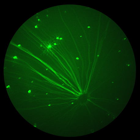
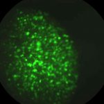
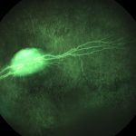
Image rétinienne fluorescente de cellules ganglionnaires marquées par la GFP, capturée avec le système MICRON, montrant des corps cellulaires brillants et discrets dispersés dans le champ rétinien.

Image rétinienne fluorescente de cellules ganglionnaires marquées par la GFP, capturée avec le système MICRON, montrant des corps cellulaires brillants et discrets dispersés dans le champ rétinien.

Image rétinienne fluorescente de cellules ganglionnaires marquées par la GFP, capturée avec le système MICRON, montrant des corps cellulaires brillants et discrets dispersés dans le champ rétinien.

Image rétinienne fluorescente de cellules ganglionnaires marquées par la GFP, capturée avec le système MICRON, montrant des corps cellulaires brillants et discrets dispersés dans le champ rétinien.

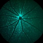
Image rétinienne de l'expression de la protéine fluorescente cyan (CFP) capturée avec le système MICRON, montrant le signal fluorescent distribué dans la rétine et le long du système vasculaire.

Image rétinienne de l'expression de la protéine fluorescente cyan (CFP) capturée avec le système MICRON, montrant le signal fluorescent distribué dans la rétine et le long du système vasculaire.

Image rétinienne de l'expression de la protéine fluorescente cyan (CFP) capturée avec le système MICRON, montrant le signal fluorescent distribué dans la rétine et le long du système vasculaire.

Image rétinienne de l'expression de la protéine fluorescente cyan (CFP) capturée avec le système MICRON, montrant le signal fluorescent distribué dans la rétine et le long du système vasculaire.

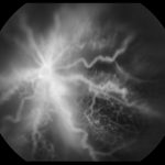
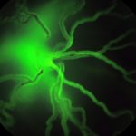
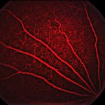
Angiographie à la fluorescéine d'une rétine de souris capturée in vivo avec le système MICRON, mettant en évidence les vaisseaux larges et fins avec un remplissage vasculaire uniforme et aucun signe de fuite ou d'occlusion. La sensibilité du système permet de visualiser le flux sanguin, les cellules individuelles étant souvent visibles en mouvement pendant la capture vidéo en direct.

Angiographie à la fluorescéine d'une rétine de souris capturée in vivo avec le système MICRON, mettant en évidence les vaisseaux larges et fins avec un remplissage vasculaire uniforme et aucun signe de fuite ou d'occlusion. La sensibilité du système permet de visualiser le flux sanguin, les cellules individuelles étant souvent visibles en mouvement pendant la capture vidéo en direct.

Angiographie à la fluorescéine d'une rétine de souris capturée in vivo avec le système MICRON, mettant en évidence les vaisseaux larges et fins avec un remplissage vasculaire uniforme et aucun signe de fuite ou d'occlusion. La sensibilité du système permet de visualiser le flux sanguin, les cellules individuelles étant souvent visibles en mouvement pendant la capture vidéo en direct.

Angiographie à la fluorescéine d'une rétine de souris capturée in vivo avec le système MICRON, mettant en évidence les vaisseaux larges et fins avec un remplissage vasculaire uniforme et aucun signe de fuite ou d'occlusion. La sensibilité du système permet de visualiser le flux sanguin, les cellules individuelles étant souvent visibles en mouvement pendant la capture vidéo en direct.

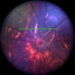
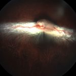
Color fundus image of a BALB/c mouse retina captured in vivo with the MICRON system, showing a translucent retina and visible choroidal vessels — features characteristic of albinism.

Color fundus image of a BALB/c mouse retina captured in vivo with the MICRON system, showing a translucent retina and visible choroidal vessels — features characteristic of albinism.

Color fundus image of a BALB/c mouse retina captured in vivo with the MICRON system, showing a translucent retina and visible choroidal vessels — features characteristic of albinism.

Color fundus image of a BALB/c mouse retina captured in vivo with the MICRON system, showing a translucent retina and visible choroidal vessels — features characteristic of albinism.
