Endless Opportunities for Your Research
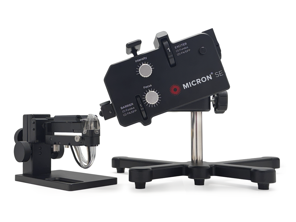
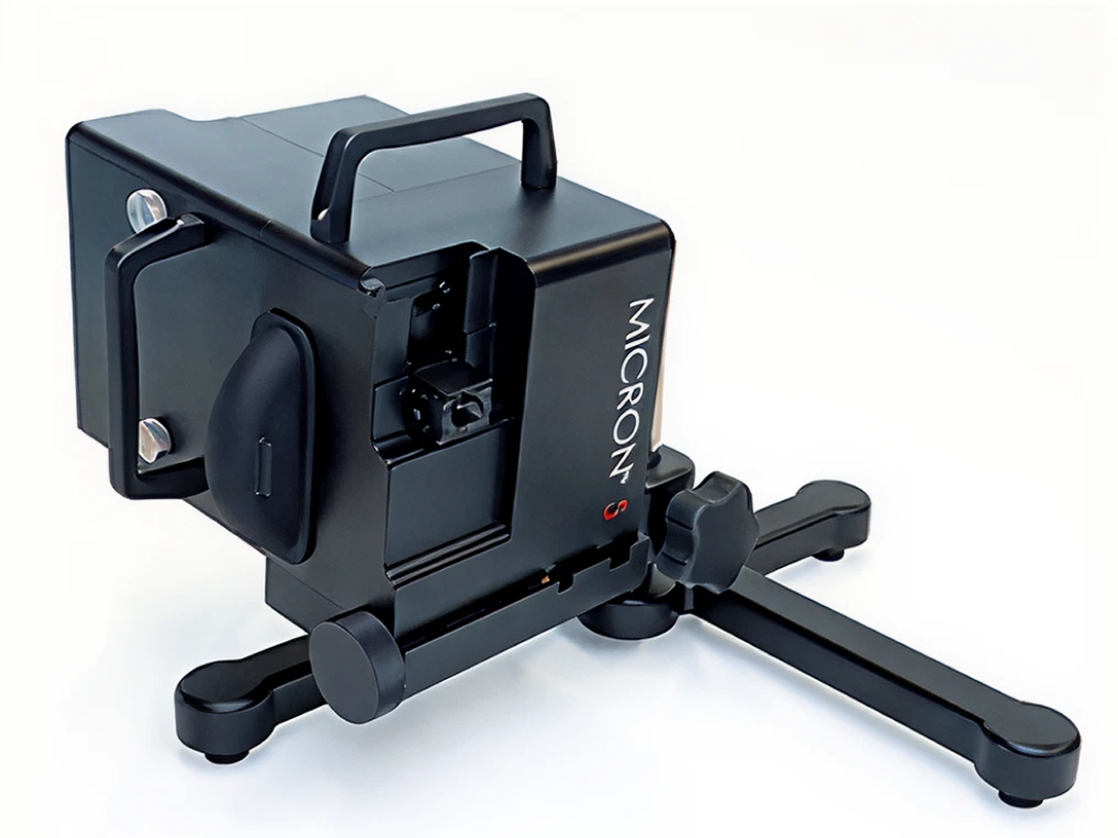
MICRON camera systems are designed to be multi-modality platforms to power your research imaging needs. See an overview of available add-on imaging modalities below.
MICRON Modalities
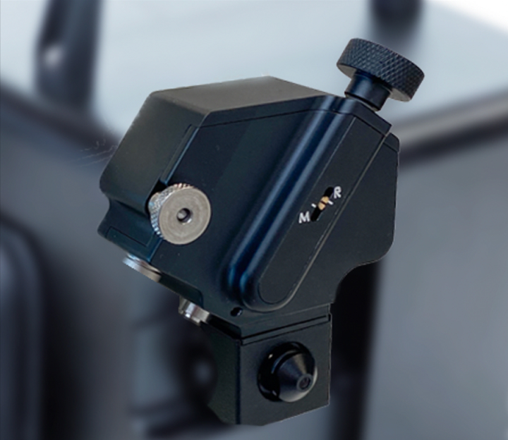
Image-guided OCT
Presenting and capturing paired color or fluorescent fundus image and OCT cross section with an axial OCT resolution below 2μ

Image-guided Lasers
Easily and precisely introduce and document laser damage models including choroidal neovascularization (CNV), retinal vein occlusion (RVO), and geographic atrophy (GA)
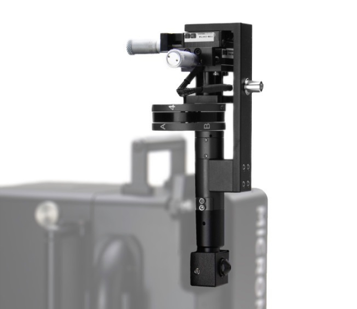
Focal ERG
Infrared light guidance for testing dark-adapted animals, to sample the ERG response of precisely located retinal positions
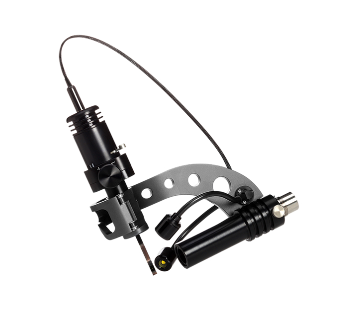
Slit Lamp
A slit lamp attachment designed for the rodent eye that delivers white light and cobalt blue anterior segment imaging with a resolution below 4μ
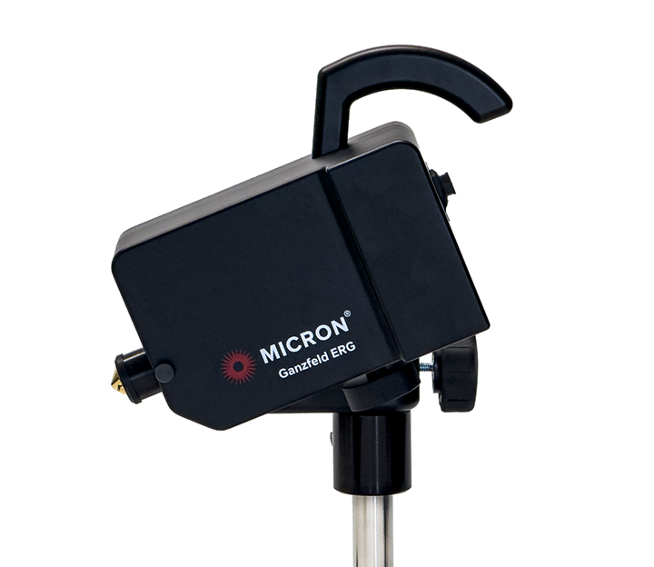
Ganzfeld ERG
Full-field ERG system optimized for the unique retinal response of rodent photoreceptors, including specific capability to excite S-Cones, M-Cones, and rods
Compatibility
All modalities are compatible with MICRON IV and MICRON 5 cameras. MICRON Image-Guided OCT2 is also compatible with MICRON SE cameras.
"Best rodent imaging system I've ever used. Produces exceptionally high quality digital fundus images of rodents and small animals. The resolution and contrast are much better than any other instrument we have tried.
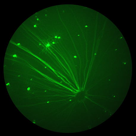
We have had good success with bright field, as well as fluorescence imaging of GFP expression in the retina and fluorescein angiography. The ability to capture video, select and output individual frames later is a big help when focusing on the retina in the small rodent eye."

John Flannery, PhD
Professor of Vision Science and Molecular and Cell Biology Associate Director, Helen Wills Neuroscience Institute University of California, Berkeley

