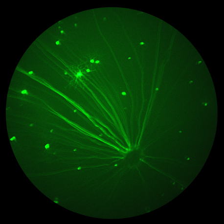
The patented MICRON 5 camera is a completely redesigned ocular imaging system developed for preclinical in vivo imaging of small animal subjects. It is designed specifically to:
- Reduce imaging session time which not only improves efficiency but also reduces the required sedation time for subject animals
- Improve data accuracy and experiment repeatability
- Capture and present more and better data for analysis
- Enhance asset utilization across groups of users
When considering the MICRON 5 ocular imaging system, especially when evaluating upgrading from MICRON IV to MICRON 5, we would like to highlight the following features and improvements:
All of these features combine to shorten the time required to image animal subjects, improve data quality and security, improve experiment repeatability, increase asset utilization through camera sharing, and more easily power complex data analysis to quickly discover research insights.
In addition, for MICRON users that take advantage of the MICRON Image-Guided OCT2 modality, Phoenix-Micron announced that it will provide automated OCT image segmentation using a new Ai / deep learning module. This will be integrated into MICRON Software Suite and ship in the Fall of 2024. The new tool will provide on-demand segmentation of OCT scans, identifying up to 10 retinal layers in an image of a healthy rodent retina. This will reduce the time and increase the accuracy of extracting key layer thickness metrics and other characteristics tied to the presence or absence of pathologies.


